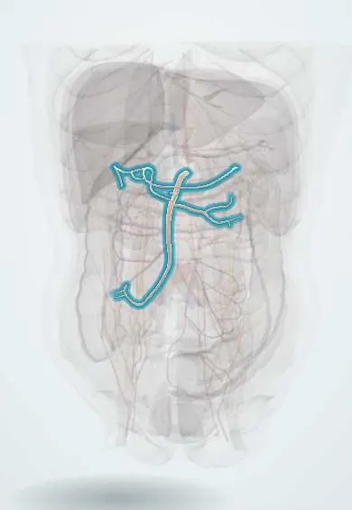
Cardiovascular system · Abdomen
Superior mesenteric artery !
The superior mesenteric artery is a large branch of the aorta that feeds the small intestine and the right half of the colon; a blockage can kill the bowel.
Opens in the Durgical app, no sign-up needed.
What it is
A large branch from the front of the aorta (at L1), running down into the mesentery.
What it does
Supplies the small intestine, the right colon and the first two-thirds of the transverse colon.
Key facts
- Origin
- Anterior aorta at L1, about 1 cm below celiac trunk; passes behind pancreatic neck, over D3 and left renal vein
- Branches
- Inferior pancreaticoduodenal, jejunal and ileal, middle colic, right colic, ileocolic
- Supplies
- Midgut: duodenum below major papilla to proximal two-thirds of transverse colon; pancreatic head
Relations
- Course
- Leaves the aorta behind the neck of the pancreas, runs down and forward over the uncinate process and the third part of the duodenum, then enters the root of the mesentery and curves toward the right iliac fossa
- In front
- Neck of the pancreas and the splenic vein above; lower down, the transverse mesocolon and the loops of the small intestine
- Behind
- From above down: aorta, left renal vein, uncinate process of the pancreas and third part of the duodenum
- Lateral
- Superior mesenteric vein, to its right
How to find it
Leaves the anterior aortic wall a short way under the celiac trunk, close to the transpyloric plane (L1). On a sagittal ultrasound it is the second vessel leaving the aorta, running down parallel to it in a collar of fat. In surgery it lies left of the superior mesenteric vein, behind the pancreatic neck and in front of the uncinate process and the third part of the duodenum.
On imaging
CT angiography (sagittal views) shows the origin, emboli as filling defects, and a narrow aortomesenteric angle or distance in superior mesenteric artery syndrome. The superior mesenteric vein twisting around the artery on CT or ultrasound (whirlpool sign) suggests midgut volvulus. Mesenteric angiography can find and treat brisk bleeding from its branches.
Common problems
- Mesenteric ischemia: a clot or narrowing starves the gut; severe pain 'out of proportion' to the examination. An emergency.
How it is diagnosed
CT angiography.
Treatment
- Restore flow (catheter or surgery) and remove dead bowel.
Clinical notes
- Acute mesenteric ischemia: an embolus, often from the heart in atrial fibrillation, or thrombosis on an atherosclerotic origin; non-occlusive ischemia occurs in low-flow states
- Chronic mesenteric ischemia (intestinal angina): pain soon after meals, fear of eating and weight loss, usually with narrowing of at least two of the three gut arteries
- Superior mesenteric artery syndrome (Wilkie syndrome): after rapid weight loss the fat around the artery shrinks, the angle narrows and the third part of the duodenum is squeezed, causing vomiting and early fullness
- Nutcracker syndrome: the left renal vein squeezed between it and the aorta can cause blood in the urine, flank pain and a left varicocele
Anatomical variants
- Replaced right hepatic artery from the superior mesenteric artery, one of the commonest hepatic variants; it runs behind the pancreatic head and portal vein and matters in pancreatic surgery
- Celiacomesenteric trunk: one common origin with the celiac trunk (rare)
- The common hepatic artery may arise from it (uncommon)
Development
The artery of the midgut, from a vitelline artery. The midgut loop herniates into the umbilical cord and rotates 270° counterclockwise around it before returning, which sets the final position of the bowel.
Related conditions
- Acute mesenteric ischaemia
- Intestinal malrotation with midgut volvulus
- Polyarteritis nodosa
- Chronic mesenteric ischemia
Test yourself
Think of the answer, then tap the question to see it.
At what level does the superior mesenteric artery arise, and what does it supply?
From the front of the aorta at L1, about 1 cm below the celiac trunk; the midgut, from the duodenum below the major papilla to the proximal two-thirds of the transverse colon.
Which structures pass between the superior mesenteric artery and the aorta?
The third part of the duodenum and the left renal vein; their compression gives superior mesenteric artery syndrome and nutcracker syndrome.
Where does an embolus usually lodge in the superior mesenteric artery?
A few centimetres past the origin, beyond the middle colic artery, sparing the upper jejunum and transverse colon; thrombosis blocks the origin and kills more bowel.
Which side of the superior mesenteric artery is the superior mesenteric vein, and what does a change suggest?
Normally on the right; a vein to the left of the artery suggests intestinal malrotation.
What is the classic presentation of acute mesenteric ischemia?
Severe abdominal pain out of proportion to the examination; it is an emergency, usually confirmed by CT angiography.
Educational information, not medical advice. For any symptom, see a health professional.
Its names
- Official Latin (TA2)
- arteria mesenterica superior; arteriae jejunales; arteriae ileales TA2 4252
- Official Spanish (DeCS)
- Arteria mesentérica superior DeCS D017538
- English (TA2)
- Superior mesenteric artery
- Also called
- SMA · arteria mesenterica cranialis
Related
Sources: Terminologia Anatomica (TA2); DeCS; Anatomy, Abdomen and Pelvis: Superior Mesenteric Artery; Anatomy of the Human Body (20th ed.): The Abdominal Aorta (Lea & Febiger (Gray's Anatomy, 1918), 1918) · adapted, Public domain.
Spotted a mistake?We are always improving for the community. Tell us what is wrong and our team will review it.Report a mistake
Representative model, for educational purposes only.