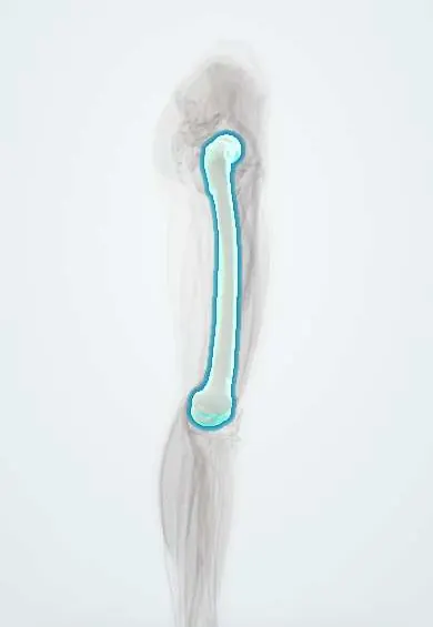
Avascular necrosis of the femoral head !
Avascular necrosis of the femoral head is death of bone in the ball of the hip after it loses its blood supply; the head can collapse.
Updated 2026-10-29
Structures involved
Tap one to see what it is and open it in 3D.
- Femur
- Femoral head cartilage
- Circumflex femoral arteries
- Ligament of the femoral head
- Hip bone
- Acetabular cartilage
- Hip capsule
In embryology
Why it happens
- Infarction: red vs white, and what decides the damage
- Fat, air and amniotic fluid embolism
- Patterns of necrosis: coagulative, liquefactive, caseous, fat, fibrinoid, gangrenous
- Bone tissue: cells, compact and spongy bone
- Ossification, growth plate and bone remodeling
What it is
Death of bone in the femoral head caused by loss of its blood supply, which can lead to collapse of the head and hip osteoarthritis. It is also called osteonecrosis.
Who gets it
Mostly adults aged about 30 to 50, often on both sides. Corticosteroid use and alcohol are the most common non-traumatic causes; sickle cell disease is an important cause in affected populations.
Causes
- Trauma: femoral neck fracture or hip dislocation
- High-dose or prolonged corticosteroid therapy
- Heavy alcohol use
- Sickle cell disease and other haemoglobinopathies
- Systemic lupus erythematosus, HIV, decompression sickness (dysbaric), Gaucher disease, radiotherapy
- Idiopathic
Risk factors
- Corticosteroid therapy
- Excess alcohol intake
- Sickle cell disease
- Lupus and other autoimmune disease
- Organ transplantation
- Smoking and clotting disorders
Pathophysiology
The femoral head is supplied mainly by the medial circumflex femoral artery with few collaterals. Trauma, fat emboli, sickled cells or raised marrow pressure cut off flow, and the bone under the cartilage dies. Repair weakens it, leading to a subchondral fracture (crescent sign), collapse and secondary arthritis.
Classification
- Ficat and Arlet stages: I normal radiograph, II sclerosis or cysts, III crescent sign and collapse, IV arthritis
- Steinberg (University of Pennsylvania) and ARCO systems add lesion size and location
Symptoms and signs
- Deep groin or buttock pain, worse with weight-bearing
- Pain that can occur at rest or at night as the disease progresses
- Limp
- Painful, limited internal rotation of the hip
- May be painless in early stages
Diagnosis
- Clinical
- Groin pain and painful internal rotation in a patient with risk factors; the other hip is also assessed.
- Tests
- Tests for the cause: blood count and haemoglobin electrophoresis, lipids, liver tests and, by context, autoimmune or HIV testing.
- Imaging
- Radiographs may be normal at first; MRI is the most sensitive test and shows the double-line sign. Later radiographs show sclerosis, the crescent sign and collapse.
Differential diagnosis
- Transient osteoporosis of the hip
- Hip osteoarthritis
- Femoral neck stress fracture
- Septic arthritis of the hip
- Lumbar radiculopathy with referred pain
- Acetabular labral tear
Goal of treatment
Preserve the femoral head before it collapses and, if it has collapsed, relieve pain and restore function, usually with hip replacement.
Treatment
- Conservative
- Protected weight-bearing, analgesia and removal of risk factors (reducing corticosteroids, stopping alcohol) in early or small lesions.
- Medical
- Bisphosphonates and other drugs have been studied for early stages; evidence is limited and practice varies.
- Surgical
- Core decompression, with or without bone grafting or cell therapy, for pre-collapse stages.
- Surgical
- Vascularised bone graft or osteotomy in selected young patients.
- Surgical
- Total hip replacement after collapse or arthritis.
Complications
- Collapse of the femoral head
- Secondary osteoarthritis of the hip
- Involvement of the other hip
- Need for hip replacement at a young age
Prognosis
Small, early lesions may stabilise, but many symptomatic lesions progress to collapse within a few years. Hip replacement gives good relief for advanced disease.
Prevention
- Using the lowest effective corticosteroid dose and duration
- Moderating alcohol intake
- Early reduction of hip dislocations and femoral neck fractures
Test yourself
Think of the answer, then tap the question to see it.
What are the main non-traumatic causes of avascular necrosis of the femoral head?
Corticosteroids and alcohol; also sickle cell disease, lupus, decompression sickness and Gaucher disease.
What is the most sensitive imaging test for early avascular necrosis of the femoral head?
MRI, which can show the double-line sign while radiographs are still normal.
What does the crescent sign mean in avascular necrosis of the femoral head?
A subchondral fracture, showing imminent or early collapse of the head (Ficat stage III).
Which artery supplies most of the femoral head in adults?
The medial circumflex femoral artery, through its retinacular branches.
What is the treatment for avascular necrosis with a collapsed femoral head and arthritis?
Total hip replacement.
Related conditions
- Hip Osteoarthritis
- Femoral Neck Fracture
- Hip dislocation
- Legg-Calvé-Perthes disease
- Slipped capital femoral epiphysis
- Systemic lupus erythematosus
- Sciatica (Lumbar Radiculopathy)
- Acetabular Labral Tear
Educational information, not medical advice. For any symptom, see a health professional.
Sources: Femoral Head Avascular Necrosis (StatPearls (NCBI Bookshelf), 2023).
Spotted a mistake?We are always improving for the community. Tell us what is wrong and our team will review it.Report a mistake
Representative model, for educational purposes only.