
Carotid-cavernous fistula
An abnormal connection between the carotid artery and the cavernous sinus: a red, bulging, sometimes pulsating eye with a bruit. It is closed endovascularly.
Quick facts
- Key signs
- Red eye with dilated · Proptosis · Double vision
- Diagnosis
- Catheter angiography
Red flags ↓
No doses: the clinician sets them by the current guideline.
What it is
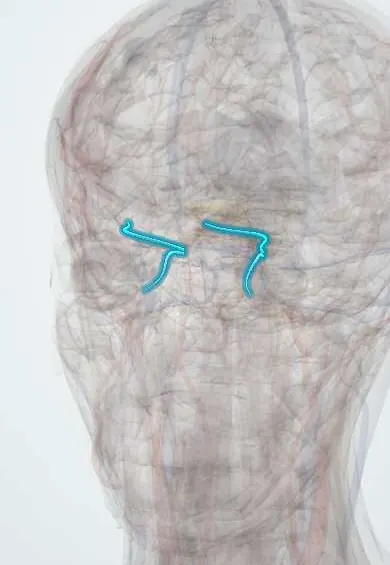
An abnormal connection between the carotid arterial system and the cavernous sinus that drives arterial blood back into the orbital veins, giving a red, swollen, proptotic eye. Direct high-flow fistulas follow head trauma and are dramatic; indirect dural low-flow fistulas arise spontaneously in older women and mimic conjunctivitis.
Who gets it
Direct fistulas mainly in young men after trauma; indirect fistulas mainly in older women, sometimes with hypertension or connective tissue disease.
Causes
- Head trauma with skull base fracture (direct)
- Rupture of an intracavernous carotid aneurysm
- Spontaneous dural shunts (indirect)
- Connective tissue disorders
Risk factors
- Head trauma
- Older age and female sex (indirect)
- Hypertension
- Connective tissue disease
Pathophysiology
High arterial pressure is transmitted to the cavernous sinus and the superior ophthalmic vein, which drains the orbit. Venous congestion swells the orbital tissues, raises eye pressure and can starve the retina and optic nerve; the nerves in the cavernous sinus can be compressed.
Classification
- Direct (high flow) or indirect (dural, low flow)
Symptoms and signs
- Red eye with dilated, tortuous ('corkscrew') conjunctival vessels
- Proptosis, sometimes pulsating
- A whooshing noise in the head
- Double vision
- Reduced vision
Diagnosis
- Clinical
- Chronic 'red eye' with corkscrew episcleral vessels, raised eye pressure and an orbital bruit on auscultation.
- Tests
- Eye pressure measurement and dilated fundus exam.
- Imaging
- CT or MR angiography show an enlarged superior ophthalmic vein and cavernous sinus; catheter angiography confirms and guides treatment.
Differential diagnosis
- Conjunctivitis
- Thyroid eye disease
- Orbital cellulitis
- Cavernous sinus thrombosis
Red eye with proptosis: which one?
| Feature | Carotid-cavernous fistula | Conjunctivitis | Thyroid eye disease | Orbital cellulitis |
|---|---|---|---|---|
| Vessels | Dilated corkscrew episcleral vessels running up to the limbus | Diffuse conjunctival redness with discharge | Redness over the muscle insertions | Diffuse redness with tense, swollen lids |
| Eye pressure | Often raised | Normal | May rise on upgaze | May rise |
| Bruit or pulsation | Present in high-flow fistulas | Absent | Absent | Absent |
| Clinical clue | Head trauma, or an older woman with spontaneous onset | Contacts, recent cold, both eyes | Thyroid disease, lid retraction | Fever, sinusitis, painful eye movements |
| Imaging | Enlarged superior ophthalmic vein and cavernous sinus | Not needed | Enlarged muscle bellies, tendons spared | Sinus disease, orbital fat stranding or abscess |
Goal of treatment
Close the fistula and protect vision.
Treatment
- Procedure
- Endovascular closure (coils or embolic material) by interventional neuroradiology.
- Medical
- Eye pressure-lowering drops while awaiting closure.
- Follow-up
- Observation of some low-flow fistulas, which can close spontaneously.
Complications
- Glaucoma
- Central retinal vein occlusion
- Cranial nerve palsies
- Loss of vision
- Intracranial haemorrhage
Common traps
- A chronic red eye that does not clear with drops is conjunctivitis: a long-standing one-sided red eye with corkscrew episcleral vessels and raised eye pressure suggests a dural fistula; listen for a bruit and image
- No bruit means no fistula: low-flow dural fistulas are often silent, so the absence of a bruit does not exclude the diagnosis
- The raised eye pressure is ordinary open-angle glaucoma: pressure rises because episcleral venous pressure is high; drops help for a while, but closing the fistula treats the cause
- Every fistula needs immediate closure: some low-flow dural fistulas close spontaneously and can be watched; falling vision, very high pressure or cortical venous drainage call for prompt treatment
- Double vision after head trauma is always a fracture or trapped muscle: the abducens nerve lies free in the cavernous sinus and is often compressed by a fistula, so look for proptosis and chemosis too
Prognosis
Good after successful closure; delayed treatment risks permanent visual loss.
Test yourself
Think of the answer, then tap the question to see it.
A young man has pulsating proptosis and a whooshing sound in his head two weeks after a motorcycle crash. What is the diagnosis and the next step?
A direct carotid-cavernous fistula; CT or MR angiography, then catheter angiography to confirm it and close it endovascularly.
Why does a carotid-cavernous fistula raise eye pressure?
Arterial blood raises pressure in the orbital and episcleral veins, so aqueous humour drains against a higher back-pressure and eye pressure rises.
Why is the abducens nerve the cranial nerve most often affected?
It runs free inside the cavernous sinus beside the carotid artery, whereas the third, fourth and ophthalmic nerves are protected in the sinus wall.
Which imaging finding makes a dural fistula urgent even when the eye looks mild?
Cortical venous drainage, because it raises the risk of intracranial haemorrhage and venous infarction.
In ophthalmology
In neurology
Connections
Educational information, not medical advice. For any symptom, see a health professional.
Sources: Carotid Cavernous Fistula (StatPearls Publishing (NCBI Bookshelf), 2023).
Spotted a mistake?We are always improving for the community. Tell us what is wrong and our team will review it.Report a mistake
Representative model, for educational purposes only.