Conditions · Ophthalmology and ENT
Thyroid eye disease (Graves orbitopathy) !
An autoimmune swelling of the tissues behind the eye, usually with Graves disease, that makes the eyes bulge and the lids retract and can threaten sight.
Updated 2026-11-01
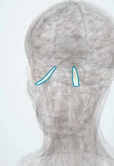
Structures involved
Tap one to see what it is and open it in 3D.
Why it happens
- Chronic inflammation: causes, cells and features
- Glycosaminoglycans (mucopolysaccharides)
- Conjunctiva, eyelid glands and lacrimal gland
What it is
An autoimmune inflammation of the orbital fat and extraocular muscles, most often linked to Graves disease. It pushes the eye forward, retracts the eyelids and can limit eye movement or compress the optic nerve.
Who gets it
The commonest cause of bulging eyes (proptosis) in adults, in either one or both eyes. It affects women several times more often than men, but severe disease is relatively more frequent in men and older people. Smokers have a much higher risk and worse disease.
Causes
- Autoantibodies against the TSH receptor (and the IGF-1 receptor) on orbital fibroblasts
- About nine in ten cases occur with Graves disease; a minority with Hashimoto thyroiditis or normal thyroid function
- Can start before, with or after the thyroid disease
Risk factors
- Smoking, the strongest modifiable risk factor
- Radioactive iodine treatment, especially in smokers or with active eye disease
- Uncontrolled hyper- or hypothyroidism
- High TSH-receptor antibody levels
- Female sex and middle age
Pathophysiology
Orbital fibroblasts carry the TSH receptor and are activated by autoantibodies and T cells. They make glycosaminoglycans that draw in water and they turn into fat cells, so the extraocular muscle bellies and orbital fat swell inside a bony box that cannot expand. The eye is pushed forward, venous drainage is impaired and, at the orbital apex, the swollen muscles can squeeze the optic nerve. The disease has an active inflammatory phase that burns out over months to a few years and leaves fibrosis.
Classification
- Activity: active (inflammatory) versus inactive (fibrotic), often scored with the clinical activity score
- Severity (EUGOGO): mild, moderate to severe, and sight-threatening (optic neuropathy or corneal breakdown)
Symptoms and signs
- Gritty, dry or watery eyes and sensitivity to light
- Bulging eyes (proptosis) and a staring look from upper lid retraction
- Lid lag on looking down
- Red, swollen eyelids and conjunctiva
- Pain behind the eyes or on eye movement
- Double vision, most often looking up, from restricted muscles
Diagnosis
- Clinical
- Typical eye signs (lid retraction, proptosis, restricted motility) with thyroid disease. Measure visual acuity, colour vision, pupils, eye movements and proptosis (exophthalmometry), and grade activity and severity.
- Tests
- TSH, free T4 and T3, and TSH-receptor antibodies.
- Imaging
- CT or MRI of the orbits shows enlarged extraocular muscle bellies with sparing of the tendons; MRI helps judge activity and CT shows the bone before decompression.
Differential diagnosis
- Orbital cellulitis
- Orbital tumour or lymphoma
- Idiopathic orbital inflammation (orbital pseudotumour)
- Carotid-cavernous fistula
- Ocular myasthenia gravis (double vision and ptosis without proptosis)
Goal of treatment
Protect sight (optic nerve and cornea), restore normal thyroid function, stop smoking, shorten the active phase and later correct the remaining changes in appearance and eye movement.
Treatment
- Lifestyle
- Stopping smoking is essential; it lowers the risk of progression and improves the response to treatment.
- Medical
- Control of thyroid function with antithyroid drugs; when radioactive iodine is used in people at risk, a short course of glucocorticoids is often given to prevent worsening of the eyes.
- Conservative
- Lubricating drops and gels, sunglasses, taping the lids at night and sleeping with the head raised; selenium is used in mild disease in some settings.
- Medical
- Moderate to severe active disease is treated with intravenous glucocorticoids; other options include immunosuppressants, biologics such as IGF-1 receptor or IL-6 blockers, and orbital radiotherapy.
- Surgical
- Urgent orbital decompression for optic neuropathy not responding to steroids. Rehabilitative surgery in the inactive phase follows a fixed order: decompression, then squint surgery, then eyelid surgery.
Complications
- Compressive optic neuropathy and permanent vision loss
- Exposure keratopathy, corneal ulcer and perforation
- Persistent double vision (restrictive strabismus)
- Lasting change in facial appearance, with a heavy psychosocial burden
Prognosis
Most cases are mild and settle over one to two years; the active phase follows a curve of worsening, plateau and improvement. A minority develop sight-threatening disease, and many keep some proptosis or lid retraction that may need surgery once inactive.
Prevention
- Not smoking, and stopping smoking in people with Graves disease
- Prompt control of thyroid function and avoiding swings into hypothyroidism after treatment
- Steroid cover with radioactive iodine in people at risk
Test yourself
Think of the answer, then tap the question to see it.
Which autoantigen on orbital fibroblasts drives thyroid eye disease?
The TSH receptor, targeted by the same autoantibodies that cause Graves hyperthyroidism (the IGF-1 receptor also takes part).
What is the most important modifiable risk factor for thyroid eye disease?
Smoking, which raises both the risk and the severity and lowers the response to treatment.
Which complication of thyroid eye disease is a sight-threatening emergency?
Compressive optic neuropathy (loss of acuity or colour vision, afferent pupillary defect); severe exposure keratopathy is the other.
What does orbital CT typically show in thyroid eye disease?
Enlarged extraocular muscle bellies with sparing of their tendons, often both eyes.
In what order is rehabilitative surgery done once the disease is inactive?
Orbital decompression first, then strabismus surgery, then eyelid surgery.
Related conditions
- Graves disease (hyperthyroidism)
- Hypothyroidism
- Thyroiditis (subacute, painless and postpartum)
- Orbital cellulitis
- Optic neuritis
- Myasthenia gravis
Educational information, not medical advice. For any symptom, see a health professional.
Sources: An overview of thyroid eye disease (Eye and Vision, 2014) · adapted, CC BY 4.0.
Spotted a mistake?We are always improving for the community. Tell us what is wrong and our team will review it.Report a mistake
Representative model, for educational purposes only.