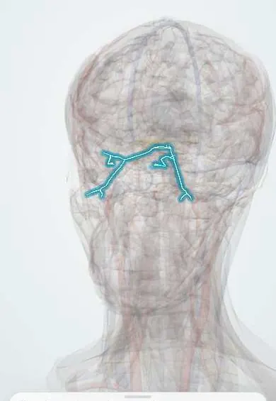
Oculomotor (third) nerve palsy !
Damage to the third cranial nerve makes the eyelid droop and the eye turn down and out, causing double vision. A blown pupil can signal an aneurysm.
Updated 2026-11-01
Structures involved
Tap one to see what it is and open it in 3D.
- Oculomotor nerve (III)
- Levator palpebrae
- Posterior communicating artery
- Posterior cerebral artery
- Internal carotid artery
- Midbrain
- Cavernous sinus
- Superior rectus
- Inferior rectus
- Medial rectus
- Inferior oblique
- Iris
Why it happens
What it is
Weakness or paralysis of the muscles supplied by the oculomotor nerve (cranial nerve III), which lifts the eyelid, moves the eye up, down and inward, and constricts the pupil. It may be complete or partial and may spare or involve the pupil.
Who gets it
One of the most frequent acquired cranial nerve palsies in adults. In older adults with diabetes or hypertension, microvascular ischaemia is the leading cause; in Colombia and Latin America, where diabetes and hypertension are common, this pattern is often seen in clinics. Compressive causes, above all aneurysms of the posterior communicating artery, are less common but are the ones that must not be missed.
Causes
- Microvascular ischaemia of the nerve (diabetes, hypertension, dyslipidaemia), usually pupil-sparing
- Compression by an aneurysm, classically at the junction of the internal carotid and posterior communicating arteries
- Uncal (transtentorial) herniation from a mass or bleed raising intracranial pressure
- Midbrain stroke, tumours, demyelination or trauma
- Cavernous sinus or orbital apex lesions (thrombosis, tumours, pituitary apoplexy), often with other cranial nerves involved
- Inflammatory causes such as giant cell arteritis, and rarely ophthalmoplegic migraine in children
Risk factors
- Diabetes mellitus
- Arterial hypertension and dyslipidaemia
- Age over 50 years
- Known or familial intracranial aneurysm, polycystic kidney disease or connective tissue disorders
- Head trauma
Pathophysiology
The pupillomotor parasympathetic fibres run on the outer (superficial) part of the nerve and get their blood from pial vessels, while the motor fibres lie deeper. An external mass such as an aneurysm compresses the outer fibres first, so the pupil dilates early. Microvascular ischaemia damages the core of the nerve and typically leaves the pupil spared. Loss of the medial, superior and inferior rectus and inferior oblique leaves the lateral rectus (VI) and superior oblique (IV) unopposed, so the eye rests down and out.
Classification
- Complete vs partial palsy
- Pupil-involving vs pupil-sparing
- By site: nuclear (midbrain), fascicular, subarachnoid, cavernous sinus, orbital
- Isolated vs with other cranial nerve or long-tract signs
Symptoms and signs
- Drooping of the upper eyelid (ptosis), often complete
- Eye turned down and out at rest
- Double vision, mainly horizontal or oblique, that disappears when the eyelid covers the eye
- Inability to move the eye inward, upward or downward
- Dilated pupil that reacts poorly to light (in compressive causes)
- Pain around the eye or forehead, which can occur in both ischaemic and aneurysmal cases
Diagnosis
- Clinical
- Inspection of the eyelid, eye position and pupils in light and dark, versions in all directions of gaze, and checking of trochlear function (intorsion on attempted downgaze) and of the other cranial nerves. The key bedside question is whether the pupil is involved.
- Tests
- Glucose, HbA1c, lipids and blood pressure for vascular risk; ESR and CRP in older adults to look for giant cell arteritis; lumbar puncture when subarachnoid haemorrhage, infection or malignancy is suspected and imaging is negative.
- Imaging
- Urgent CT angiography or MR angiography when the pupil is involved, the palsy is partial or there are other worrying features; catheter angiography if non-invasive imaging is inconclusive. MRI of the brain and orbits looks for midbrain, cavernous sinus and orbital lesions.
Differential diagnosis
- Myasthenia gravis (fluctuating ptosis and ophthalmoplegia with a normal pupil)
- Horner syndrome (mild ptosis with a small pupil)
- Internuclear ophthalmoplegia
- Thyroid eye disease and orbital disease
- Fourth or sixth nerve palsy and skew deviation
- Pharmacological mydriasis
Goal of treatment
Find and treat the cause promptly, above all an aneurysm or herniation, relieve double vision and protect the eye while the nerve recovers.
Treatment
- Acute
- A pupil-involving or otherwise suspicious palsy is a neurosurgical emergency: an aneurysm is treated by endovascular coiling or surgical clipping, and raised intracranial pressure is managed urgently.
- Medical
- For microvascular palsy, control of glucose, blood pressure and lipids, with antiplatelet therapy in selected patients; corticosteroids when giant cell arteritis or another inflammatory cause is confirmed.
- Conservative
- Occlusion of one eye (patch or occlusive lens) or temporary prisms to relieve double vision while recovery takes place.
- Follow-up
- Close follow-up of the pupil and eye movements in the first days and weeks; any new pupil involvement or other neurological sign leads to urgent vascular imaging.
- Surgical
- Strabismus surgery or eyelid surgery for stable deficits that persist after six to twelve months.
Complications
- Rupture of an underlying aneurysm with subarachnoid haemorrhage
- Permanent ptosis or strabismus with lasting double vision
- Aberrant regeneration (for example, eyelid elevation on attempted downgaze or adduction), which suggests compression or trauma rather than ischaemia
- Amblyopia in young children
Prognosis
Microvascular palsies usually recover partly or completely within about three to six months. Outcomes in compressive and traumatic cases depend on the cause and on how quickly it is treated; aneurysmal palsies may improve after the aneurysm is secured.
Test yourself
Think of the answer, then tap the question to see it.
What is the classic eye position in a complete oculomotor nerve palsy?
Down and out, with ptosis, because the lateral rectus and superior oblique act unopposed.
Why does an aneurysm usually dilate the pupil while a diabetic palsy spares it?
Pupillary fibres run superficially and are compressed first by an external mass, whereas ischaemia damages the core of the nerve.
Which aneurysm classically causes a painful pupil-involving third nerve palsy?
An aneurysm of the posterior communicating artery at its junction with the internal carotid artery.
What is the first imaging step for a pupil-involving third nerve palsy?
Urgent CT angiography or MR angiography of the intracranial vessels.
A drowsy patient with a fixed dilated pupil after a head injury most likely has what?
Uncal (transtentorial) herniation compressing the oculomotor nerve, a neurosurgical emergency.
Related conditions
- Subarachnoid haemorrhage
- Type 2 diabetes mellitus
- Type 1 diabetes mellitus
- Horner syndrome
- Myasthenia gravis
- Raised intracranial pressure and brain herniation
- Giant cell arteritis
- Brainstem stroke syndromes (Wallenberg and others)
- Pituitary adenoma
- Migraine
Educational information, not medical advice. For any symptom, see a health professional.
Sources: Cranial mononeuropathy III - diabetic type: MedlinePlus Medical Encyclopedia (MedlinePlus (U.S. National Library of Medicine), 2025).
Spotted a mistake?We are always improving for the community. Tell us what is wrong and our team will review it.Report a mistake
Representative model, for educational purposes only.