Conditions · Nephrology and urology
CKD-mineral and bone disorder (renal osteodystrophy) !
In chronic kidney disease the balance of calcium, phosphate, vitamin D and parathyroid hormone breaks down, weakening bones and hardening blood vessels.
Updated 2026-11-01
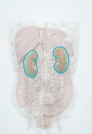
Structures involved
Tap one to see what it is and open it in 3D.
In the clinical exam
Why it happens
- Pathologic calcification: dystrophic vs metastatic
- Hyperplasia: physiologic and pathologic
- Calcium regulation: PTH, vitamin D and calcitonin
- Phosphate and FGF-23
- Bone remodeling
- Thyroid follicles, C cells and parathyroid
What it is
A systemic disorder of chronic kidney disease made up of abnormal calcium, phosphate, parathyroid hormone (PTH) and vitamin D levels, abnormal bone (renal osteodystrophy) and calcification of vessels and soft tissues.
Who gets it
Develops in almost everyone whose chronic kidney disease reaches the advanced stages and is nearly universal in people on dialysis. In Colombia and Latin America, where diabetes and hypertension drive a growing dialysis population, it is a frequent cause of fractures, bone pain and cardiovascular disease in renal units.
Causes
- Falling kidney excretion of phosphate as glomerular filtration declines
- Reduced kidney activation of vitamin D (less calcitriol)
- Rising FGF23 and, later, parathyroid hyperplasia with secondary hyperparathyroidism
- Factors from treatment: calcium load, aluminium exposure in the past, glucocorticoids after transplant
Risk factors
- Advanced chronic kidney disease (stages 3b–5) and dialysis
- Diabetes and hypertension as the underlying kidney disease
- High dietary phosphate, especially from processed food additives
- Poor adherence to diet, dialysis or phosphate binders
- Long duration on dialysis
Pathophysiology
As nephrons are lost, phosphate is retained and FGF23 rises, which further suppresses calcitriol. Low calcitriol and low calcium with high phosphate stimulate the parathyroid glands, which enlarge and secrete more PTH (secondary hyperparathyroidism). High PTH drives high-turnover bone disease (osteitis fibrosa cystica); oversuppression causes low-turnover adynamic bone. The calcium–phosphate excess deposits in arteries and soft tissue. With long-standing stimulation the glands may become autonomous (tertiary hyperparathyroidism) with high calcium.
Classification
- Secondary hyperparathyroidism: high PTH with low or normal calcium and high phosphate
- Tertiary hyperparathyroidism: autonomous PTH secretion with high calcium, often after transplant
- High-turnover bone disease (osteitis fibrosa cystica)
- Low-turnover disease: adynamic bone disease and osteomalacia
- Mixed uraemic osteodystrophy
Symptoms and signs
- Often silent for years; detected on routine blood tests
- Bone and joint pain, proximal muscle weakness
- Fractures with little trauma, including hip fractures
- Pruritus (itching), worse with high phosphate and PTH
- In children: growth failure and bone deformities (renal rickets)
- Signs of vascular calcification, such as hard arteries or a calcified aorta on X-ray
Diagnosis
- Clinical
- Suspected in every patient with chronic kidney disease from stage 3 onward; history of fractures, bone pain, itching and medications, plus examination of the skin and bones.
- Tests
- Serial serum calcium, phosphate, intact PTH and alkaline phosphatase, with 25-hydroxyvitamin D. Trends matter more than single values. Bone biopsy with tetracycline labelling is the reference test for bone turnover but is rarely needed.
- Imaging
- Plain films may show subperiosteal resorption of the phalanges, a salt-and-pepper skull or a rugger-jersey spine; lateral abdominal X-ray or echocardiography to detect vascular and valve calcification; bone density scanning to estimate fracture risk.
Differential diagnosis
- Primary hyperparathyroidism (high calcium with normal kidney function)
- Vitamin D deficiency osteomalacia without kidney disease
- Postmenopausal or age-related osteoporosis
- Bone metastases or multiple myeloma
- Dialysis-related amyloidosis
Goal of treatment
Bring phosphate toward normal, avoid both hypo- and hypercalcaemia, keep PTH within a target range for the stage of disease, and protect bones and blood vessels.
Treatment
- Lifestyle
- Diet low in phosphate, especially avoiding processed foods and drinks with phosphate additives, guided by a renal dietitian.
- Medical
- Phosphate binders taken with meals (calcium-based and non-calcium-based classes); adequate dialysis also removes phosphate.
- Medical
- Correction of vitamin D deficiency, active vitamin D (calcitriol or analogues) and calcimimetics to lower PTH in secondary hyperparathyroidism.
- Follow-up
- Regular calcium, phosphate, PTH and alkaline phosphatase at intervals set by CKD stage; nursing focus on binder timing with meals, diet teaching, itching and fall prevention.
- Surgical
- Parathyroidectomy for severe hyperparathyroidism not controlled by medicines, and in tertiary hyperparathyroidism; kidney transplantation corrects much of the disorder.
Complications
- Fractures, bone pain and skeletal deformity
- Vascular and valve calcification, raising cardiovascular death
- Calciphylaxis (calcific uraemic arteriolopathy), with painful skin necrosis
- Tertiary hyperparathyroidism with hypercalcaemia
- Growth failure in children
- Hungry bone syndrome with severe hypocalcaemia after parathyroidectomy
Prognosis
A chronic disorder that lasts as long as kidney failure. High phosphate and vascular calcification are linked to higher cardiovascular mortality; good control and timely transplantation improve bone health, though some changes persist after transplant.
Prevention
- Early detection and slowing of chronic kidney disease
- Monitoring mineral levels from stage 3 and starting diet and treatment early
Test yourself
Think of the answer, then tap the question to see it.
What laboratory pattern is typical of secondary hyperparathyroidism in chronic kidney disease?
High phosphate, low or normal calcium, low calcitriol and high PTH, with high FGF23.
Why does calcitriol fall in chronic kidney disease?
The damaged kidney makes less 1-alpha-hydroxylase activity, and high FGF23 from phosphate retention suppresses it further.
What is tertiary hyperparathyroidism?
Autonomous parathyroid hormone secretion after long-standing stimulation, causing high calcium, typically in long-term dialysis or after transplant.
What are two classic X-ray findings of renal osteodystrophy?
Subperiosteal bone resorption of the radial side of the middle phalanges, and a rugger-jersey spine (also a salt-and-pepper skull).
What is calciphylaxis?
Calcification and thrombosis of small skin arterioles in advanced kidney disease, causing very painful necrotic skin ulcers with high mortality.
Related conditions
- Chronic kidney disease
- Primary hyperparathyroidism
- Hypocalcaemia
- Hypercalcaemia
- Hyperkalaemia
- Rickets and osteomalacia
- Osteoporosis
- Diabetic kidney disease
Educational information, not medical advice. For any symptom, see a health professional.
Sources: Mineral & Bone Disorder in Chronic Kidney Disease - NIDDK (National Institute of Diabetes and Digestive and Kidney Diseases (NIDDK), 2024); CKD-Mineral and Bone Disorder (CKD-MBD) – KDIGO (KDIGO, 2017); Chronic kidney disease: MedlinePlus Medical Encyclopedia (MedlinePlus (U.S. National Library of Medicine), 2024).
Spotted a mistake?We are always improving for the community. Tell us what is wrong and our team will review it.Report a mistake
Representative model, for educational purposes only.