Endocrine system · Head and neck
Parathyroid glands !
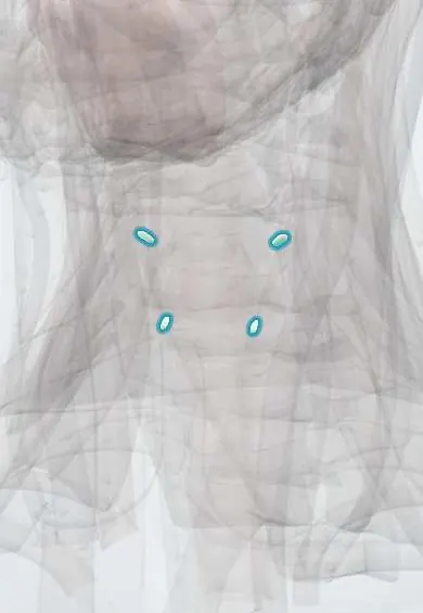
The parathyroid glands are usually four rice-grain-sized glands on the back of the thyroid; they control the level of calcium in the blood.
Opens in the Durgical app, no sign-up needed.
What it is
Usually four rice-grain-sized glands on the back of the thyroid.
What it does
Control calcium in the blood.
Key facts
- Where
- Back of the thyroid lobes (usually 4)
- Blood supply
- Inferior thyroid artery (mainly)
- Nerve
- Controlled by blood calcium
- Function
- Parathyroid hormone raises blood calcium
Relations
- In front
- Back of the thyroid gland lobes, inside or just outside the thyroid capsule
- Behind
- Tracheo-oesophageal groove: the superior glands usually lie behind (dorsal to) the recurrent laryngeal nerve, the inferior ones in front of it
- Above
- Superior glands sit near where the recurrent laryngeal nerve enters the larynx, at the cricoid cartilage level
- Below
- Inferior glands lie near the lower pole, often close to where the inferior thyroid artery enters the gland, or in the thyrothymic ligament
How to find it
Not palpable when normal. At surgery the superior glands are sought on the back of the upper thyroid lobe, near where the recurrent laryngeal nerve enters the larynx and usually deep (dorsal) to the nerve; the inferior glands lie near the lower pole, superficial (ventral) to the nerve, often beside thymic tissue.
Examination
- Chvostek sign: tapping over the facial nerve in front of the ear twitches the corner of the mouth; points to low calcium
- Trousseau sign: a blood-pressure cuff inflated above systolic pressure for a few minutes brings on a spasm of the hand; a more specific sign of low calcium
On imaging
Normal glands are usually too small to see. An overactive gland is located with ultrasound, sestamibi scintigraphy (often with SPECT/CT) or four-dimensional CT before targeted surgery.
Common problems
- Hyperparathyroidism: high calcium causing kidney stones, bone loss and tiredness.
- Can be injured in thyroid surgery, causing low calcium (tingling, cramps).
How it is diagnosed
Blood calcium and parathyroid hormone; ultrasound and nuclear scans to find an overactive gland.
Treatment
- Removing the overactive gland.
- Preserving them during thyroidectomy; calcium and vitamin D if calcium drops.
Clinical notes
- Ectopic sites include the thymus and mediastinum, the carotid sheath, inside the thyroid and behind the oesophagus; a missed ectopic adenoma is a cause of failed surgery
- In surgery they are told apart from fat and lymph nodes by their tan colour; a gland that loses its blood supply can be minced and reimplanted into a neck or forearm muscle
Anatomical variants
- More than four glands (supernumerary) in a minority, fewer than four found in some
- Inferior glands anywhere along the thymus path, from high in the neck (undescended) to the mediastinum
Development
They bud from the third and fourth pharyngeal pouches; the third-pouch glands are carried down with the thymus past the fourth-pouch glands, so the pair that ends up inferior starts higher in the embryo and has the less predictable final site.
Memory aid
Primary hyperparathyroidism: "stones, bones, abdominal groans and psychic moans" (kidney stones, bone pain and loss, abdominal pain and constipation, low mood and confusion).
Related conditions
- Hypocalcaemia
- Hypercalcaemia
- Primary hyperparathyroidism
- Hypoparathyroidism (incl. post-thyroidectomy hypocalcaemia)
- Multiple endocrine neoplasia (MEN 1, 2A, 2B)
- Autoimmune polyendocrine syndromes (APS-1, APS-2) and IPEX
- Hypercalcaemia of malignancy
- CKD-mineral and bone disorder (renal osteodystrophy)
- Hypophosphataemia
- DiGeorge syndrome (22q11.2 deletion syndrome)
Test yourself
Think of the answer, then tap the question to see it.
From which pharyngeal pouches do the superior and inferior parathyroid glands develop?
Inferior glands from the third pouch with the thymus; superior glands from the fourth. The inferior ones migrate further, so their position varies more.
Which artery mainly supplies the parathyroid glands?
The inferior thyroid artery.
What are the Chvostek and Trousseau signs, and what do they point to after thyroidectomy?
Chvostek: tapping the facial nerve twitches the mouth corner. Trousseau: an inflated cuff brings on hand spasm. Both point to low calcium from parathyroid injury.
Where can an ectopic inferior parathyroid adenoma be found?
Anywhere along the thymus path, from high in the neck to the mediastinum; also the carotid sheath, inside the thyroid or behind the oesophagus. A missed one causes failed surgery.
How do the superior and inferior parathyroid glands relate to the recurrent laryngeal nerve?
The superior glands usually lie behind (dorsal to) the recurrent laryngeal nerve; the inferior glands lie in front of it.
Educational information, not medical advice. For any symptom, see a health professional.
Its names
- Official Latin (TA2)
- glandulae parathyreoideae TA2 3870
- Official Spanish (DeCS)
- Glándulas Paratiroides DeCS D010280
- English (TA2)
- Parathyroid glands
Related
Sources: Terminologia Anatomica (TA2); DeCS; Anatomy, Head and Neck, Parathyroid; Anatomy of the Human Body (20th ed.): The Branchial Region (Lea & Febiger (Gray's Anatomy, 1918), 1918) · adapted, Public domain.
Spotted a mistake?We are always improving for the community. Tell us what is wrong and our team will review it.Report a mistake
Representative model, for educational purposes only.