Eosinophilic pneumonias (including Löffler syndrome) !
Lung inflammation where eosinophils, a type of white blood cell, fill the air spaces; causes include parasites, drugs and unknown triggers.
Updated 2026-11-01
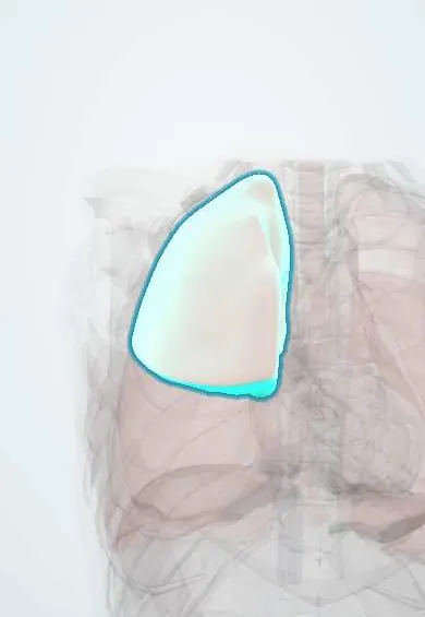
Structures involved
Tap one to see what it is and open it in 3D.
Why it happens
- White blood cells: types and functions
- Innate and adaptive immunity: overview
- The alveolus: type I and II pneumocytes, surfactant, the blood–air barrier
What it is
A group of lung diseases in which eosinophils accumulate in the alveoli and interstitium, usually with blood eosinophilia. It includes simple pulmonary eosinophilia (Löffler syndrome) from migrating parasite larvae, acute eosinophilic pneumonia, chronic eosinophilic pneumonia, drug-induced forms and tropical pulmonary eosinophilia.
Who gets it
Löffler syndrome is relevant in Colombia and Latin America, where Ascaris, hookworm and Strongyloides infections remain common in rural and poor urban areas, especially in children. Acute eosinophilic pneumonia affects young adults, often soon after starting to smoke or vape; chronic eosinophilic pneumonia affects mainly middle-aged women, many with asthma.
Causes
- Larval lung migration of Ascaris lumbricoides, hookworms (Ancylostoma, Necator) and Strongyloides stercoralis (Löffler syndrome)
- Drugs: nitrofurantoin, non-steroidal anti-inflammatories, some antibiotics, anticonvulsants and others
- New or resumed smoking, vaping and inhaled dusts (acute eosinophilic pneumonia)
- Idiopathic (chronic eosinophilic pneumonia)
- Filarial worms (tropical pulmonary eosinophilia), ABPA and eosinophilic granulomatosis with polyangiitis
Risk factors
- Poor sanitation, walking barefoot and soil contact in endemic areas
- Recent start of a new drug
- Recent start of tobacco or electronic cigarette use
- Asthma and atopy (chronic form)
- Immunosuppression with Strongyloides (risk of hyperinfection)
Pathophysiology
An antigen (larvae, drug or inhaled agent) drives a Th2 response with interleukin-5, which recruits and activates eosinophils. Eosinophils fill the alveoli and release toxic granule proteins that injure the epithelium, producing infiltrates and impaired gas exchange. In Löffler syndrome the larvae cross from the blood into the alveoli and ascend the airways, causing transient migratory infiltrates for one to two weeks before they are swallowed and mature in the gut.
Classification
- Simple pulmonary eosinophilia (Löffler syndrome): mild, transient, migratory infiltrates
- Acute eosinophilic pneumonia: days, fever and hypoxaemic respiratory failure, blood eosinophils often normal at onset
- Chronic eosinophilic pneumonia: weeks to months, peripheral infiltrates, blood eosinophilia
- Secondary: drug-induced, parasitic (including tropical pulmonary eosinophilia), ABPA, vasculitis
Symptoms and signs
- Dry cough and wheeze
- Fever and malaise
- Breathlessness, severe in the acute form
- Night sweats and weight loss in the chronic form
- Few or no symptoms in Löffler syndrome, sometimes with urticaria
Diagnosis
- Clinical
- Suspected when pulmonary infiltrates occur with blood or lung eosinophilia. The history focuses on drugs, smoking or vaping, travel, soil exposure and asthma. Strongyloides infection must be excluded before glucocorticoids are started.
- Tests
- Full blood count with eosinophil count; total IgE. Bronchoalveolar lavage with eosinophils above 25% confirms acute eosinophilic pneumonia. Stool examination for ova and larvae (often negative during the migratory phase, positive weeks later) and Strongyloides serology. ANCA and other tests when vasculitis is suspected.
- Imaging
- Chest X-ray: migratory, fleeting infiltrates in Löffler syndrome; peripheral infiltrates sparing the centre («photographic negative of pulmonary oedema») in chronic eosinophilic pneumonia. CT: ground-glass opacities and consolidation; diffuse infiltrates with small effusions in the acute form.
Differential diagnosis
- Community-acquired pneumonia
- ARDS and acute interstitial pneumonia
- Allergic bronchopulmonary aspergillosis
- Eosinophilic granulomatosis with polyangiitis
- Cryptogenic organising pneumonia and hypersensitivity pneumonitis
Goal of treatment
Identify and remove the cause, treat parasites, suppress eosinophilic inflammation when needed and support oxygenation in severe forms.
Treatment
- Conservative
- Löffler syndrome is usually self-limited; withdrawal of the suspected drug and smoking or vaping cessation are essential in secondary forms.
- Medical
- Antiparasitic therapy (albendazole, mebendazole, ivermectin for Strongyloides) once parasites are confirmed or suspected.
- Medical
- Systemic glucocorticoids for acute and chronic eosinophilic pneumonia, with rapid response; chronic forms often relapse when tapered.
- Acute
- Oxygen and ventilatory support in acute eosinophilic pneumonia with respiratory failure.
Complications
- Acute hypoxaemic respiratory failure
- Relapse of chronic eosinophilic pneumonia
- Strongyloides hyperinfection when glucocorticoids are given without screening
- Adverse effects of prolonged glucocorticoids
Prognosis
Löffler syndrome resolves in days to weeks. Acute eosinophilic pneumonia responds dramatically to glucocorticoids and rarely recurs if the trigger is avoided. Chronic eosinophilic pneumonia responds well but often relapses.
Prevention
- Sanitation, safe water, footwear and periodic deworming in endemic areas
- Avoiding tobacco and electronic cigarettes
- Strongyloides screening or empirical ivermectin before immunosuppression in at-risk people
Test yourself
Think of the answer, then tap the question to see it.
What causes Löffler syndrome?
Transpulmonary migration of helminth larvae, mainly Ascaris, hookworms and Strongyloides, producing transient migratory infiltrates with eosinophilia.
Why can stool examination be negative early in Löffler syndrome?
Larvae are still migrating through the lungs and adult worms have not yet produced eggs in the gut.
Which radiological pattern suggests chronic eosinophilic pneumonia?
Peripheral infiltrates sparing the central lung, the «photographic negative of pulmonary oedema».
What bronchoalveolar lavage finding supports acute eosinophilic pneumonia?
Eosinophils above 25% of lavage cells.
What must be excluded before giving glucocorticoids?
Strongyloides infection, because steroids can precipitate fatal hyperinfection.
Related conditions
- Soil-transmitted helminthiasis (ascariasis, hookworm, trichuriasis)
- Strongyloidiasis
- Toxocariasis (visceral and ocular larva migrans)
- Allergic bronchopulmonary aspergillosis (ABPA)
- Eosinophilic granulomatosis with polyangiitis (Churg–Strauss)
- Asthma
- Acute respiratory distress syndrome (ARDS)
- Cutaneous drug reactions and DRESS
- Community-acquired pneumonia
Educational information, not medical advice. For any symptom, see a health professional.
Sources: Simple pulmonary eosinophilia (MedlinePlus Medical Encyclopedia (U.S. National Library of Medicine), 2024); Eosinophilic Pneumonia (National Jewish Health, 2024).
Spotted a mistake?We are always improving for the community. Tell us what is wrong and our team will review it.Report a mistake
Representative model, for educational purposes only.