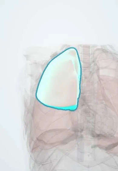
Right upper lobe !
The right upper lobe is the top part of the right lung, in the upper right chest; it is a common site for tuberculosis and lung cancer.
Official name: Superior lobe of right lung
Opens in the Durgical app, no sign-up needed.
What it is
The top lobe of the right lung, above the horizontal fissure.
What it does
Fills the upper right chest; a common site for tuberculosis and lung cancer.
Key facts
- Where
- Above horizontal and oblique fissures; segments apical, posterior, anterior
- Blood supply
- Pulmonary artery branches (gas exchange); bronchial arteries (nourish lung tissue)
- Nerve
- Pulmonary plexus: vagus (bronchoconstriction, secretion); upper thoracic sympathetic (bronchodilation)
- Function
- Gas exchange; posterior segment is a classic aspiration site when supine
Relations
- Above
- Cervical pleura and suprapleural membrane; above them the subclavian artery, the lower trunk of the brachial plexus and the stellate ganglion
- Medial
- Superior vena cava, right brachiocephalic vein, trachea, right vagus nerve and the arch of the azygos vein, which grooves the lobe above the hilum
- Below
- Right middle lobe across the horizontal fissure in front; right lower lobe across the oblique fissure behind
How to find it
The apex is heard above the medial third of the clavicle. In front, the lobe lies above the 4th costal cartilage (horizontal fissure); behind, only above about the T3 spinous process, where the oblique fissure begins. Its sounds are best heard on the upper front of the chest and high in the axilla.
On imaging
Consolidation is bounded below by a sharp horizontal fissure. Collapse lifts the fissure and the right hilum. Apical lesions can hide behind the clavicle and first rib; an apical (lordotic) view or CT shows them. Cavities with upper lobe nodules suggest active tuberculosis.
Common problems
- Pneumonia (infection), asthma and COPD (narrowed airways).
- Lung cancer, often linked to smoking.
- Pulmonary embolism: a clot blocks an artery in the lung.
How it is diagnosed
Listening to the chest, chest X-ray, CT, breathing tests (spirometry), blood oxygen.
Treatment
- Depends on the cause: antibiotics, inhalers, oxygen; surgery removes a lobe or segment for some cancers.
Clinical notes
- Tuberculosis remains a public health problem in Colombia, which keeps upper lobe cavitation high on the list of causes
- Pancoast (superior sulcus) tumour: shoulder and ulnar-side arm pain and hand weakness from the brachial plexus, and Horner syndrome from the sympathetic chain
- Aspiration while lying on the right side tends to reach the posterior segment of this lobe
Anatomical variants
- Azygos lobe: the azygos vein runs through the apex inside a fold of pleura, creating an extra fissure; uncommon, seen as a fine curved line at the right apex on X-ray
Development
Around week 5 the lung buds split into lobar buds: three on the right and two on the left, which is why the left lung lacks a middle lobe. On the right, the upper of the three buds forms this lobe.
Related conditions
- Tuberculosis
- Lambert-Eaton myasthenic syndrome
- Cystic fibrosis
- Sarcoidosis
- Pneumoconioses (silicosis, asbestosis, coal workers' pneumoconiosis)
- Solitary pulmonary nodule
- Hypersensitivity pneumonitis
- Eosinophilic pneumonias (including Löffler syndrome)
- Chronic obstructive pulmonary disease (COPD)
- Pneumothorax
- Pulmonary tuberculosis
- Lung cancer
Test yourself
Think of the answer, then tap the question to see it.
Where in the right upper lobe does reactivation tuberculosis classically occur, and why?
In the apical and posterior segments, where oxygen tension is highest.
What are the features of a Pancoast tumour at the apex of the right upper lobe?
Shoulder and ulnar-side arm pain and hand weakness from the brachial plexus, and Horner syndrome from the sympathetic chain.
What is the Golden S sign?
Right upper lobe collapse with a central mass: the horizontal fissure is pulled up and in, and its edge forms a reversed S.
What is an azygos lobe?
An uncommon variant: the azygos vein runs through the right apex inside a fold of pleura, seen as a fine curved line on X-ray.
Educational information, not medical advice. For any symptom, see a health professional.
Its names
- Official Latin (TA2)
- lobus superior pulmonis dextri TA2 3284
- Official Spanish (DeCS)
- Lóbulo superior del pulmón derecho DeCS D008168
- English (TA2)
- Superior lobe of right lung
- Also called
- upper lobe of right lung · lobus ventrocranialis
Sources: Terminologia Anatomica (TA2); DeCS; Anatomy, Thorax, Lungs; 22.7 Embryonic Development of the Respiratory System (OpenStax (Rice University), 2022) · adapted, CC BY 4.0; Anatomy of the Human Body (20th ed.): The Respiratory Apparatus (Lea & Febiger (Gray's Anatomy, 1918), 1918) · adapted, Public domain.
Spotted a mistake?We are always improving for the community. Tell us what is wrong and our team will review it.Report a mistake
Representative model, for educational purposes only.