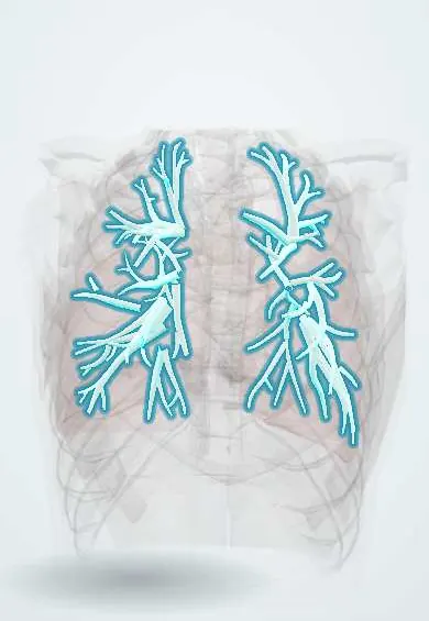
Allergic bronchopulmonary aspergillosis (ABPA) !
An allergic reaction to the mould Aspergillus in the airways of people with asthma or cystic fibrosis that can scar and widen the bronchi.
Updated 2026-11-01
Structures involved
Tap one to see what it is and open it in 3D.
Why it happens
- Cystic fibrosis: from the CFTR channel to organ damage
- Innate and adaptive immunity: overview
- Airway defenses, cough and mucociliary clearance
What it is
A hypersensitivity lung disease in which Aspergillus fumigatus colonises the bronchi of patients with asthma or cystic fibrosis and triggers an intense allergic (IgE) and eosinophilic response. It is not an invasive infection; invasive aspergillosis and aspergilloma are on the aspergillosis page.
Who gets it
Affects an estimated 1 to 3% of adults with asthma (more in severe asthma) and a larger share of people with cystic fibrosis. It is underdiagnosed in Latin America because total IgE and Aspergillus-specific tests are not always requested; it should be sought in every patient with difficult-to-control asthma.
Causes
- Hypersensitivity to Aspergillus fumigatus, a mould that is everywhere in soil, compost and indoor dust
- Underlying asthma or cystic fibrosis that allows spores to persist in thick bronchial mucus
- Genetic predisposition (HLA class II variants, CFTR variants)
Risk factors
- Severe or poorly controlled asthma
- Cystic fibrosis
- Atopy and high environmental mould exposure
- Long-standing bronchiectasis
Pathophysiology
Inhaled spores germinate in the mucus of abnormal airways and release antigens and proteases. A Th2-dominant response produces large amounts of total and Aspergillus-specific IgE and IgG, and recruits eosinophils (type I and type III hypersensitivity). Eosinophil-rich mucus plugs obstruct segmental bronchi, causing atelectasis and fleeting infiltrates; repeated inflammation damages the bronchial wall and produces central (proximal) bronchiectasis, which may end in upper-lobe fibrosis.
Classification
- Serological ABPA (without bronchiectasis)
- ABPA with bronchiectasis
- ABPA with high-attenuation mucus on CT
- Clinical stages: acute, response, remission, exacerbation, treatment-dependent, advanced fibrotic disease
Symptoms and signs
- Worsening asthma with wheeze that resists usual inhaled treatment
- Productive cough with brownish mucus plugs or casts
- Low-grade fever, malaise and weight loss during flares
- Pleuritic chest pain
- Haemoptysis in advanced bronchiectasis
Diagnosis
- Clinical
- Considered in every patient with asthma that is hard to control and in cystic fibrosis with clinical deterioration. Diagnosis combines a predisposing condition, sensitisation to Aspergillus and raised total IgE, plus supporting findings.
- Tests
- Positive Aspergillus skin prick test or raised Aspergillus fumigatus-specific IgE; total serum IgE markedly raised (usually above 500 to 1000 IU/mL). Supporting: Aspergillus-specific IgG (precipitins), peripheral eosinophilia, sputum culture. Total IgE is followed to monitor response and relapse. Spirometry shows obstruction.
- Imaging
- Chest X-ray: fleeting upper-lobe infiltrates, «finger-in-glove» opacities from mucus-filled bronchi, «tram-track» lines. High-resolution CT: central bronchiectasis, mucus plugging (high-attenuation mucus is very specific), centrilobular nodules.
Differential diagnosis
- Severe asthma without ABPA
- Chronic pulmonary aspergillosis and aspergilloma
- Eosinophilic pneumonia and eosinophilic granulomatosis with polyangiitis
- Bronchiectasis from other causes (post-tuberculosis, cystic fibrosis alone, immunodeficiency)
- Pulmonary tuberculosis
Goal of treatment
Suppress the allergic inflammation, control asthma, prevent exacerbations and stop progression to bronchiectasis and fibrosis.
Treatment
- Medical
- Oral glucocorticoids are first line for acute ABPA, tapered over months while total IgE and imaging are followed.
- Medical
- Oral azole antifungals (itraconazole, voriconazole) to reduce fungal burden and allow lower steroid exposure.
- Medical
- Biologic therapy against IgE or the interleukin-5 pathway in steroid-dependent or recurrent disease; optimised inhaled asthma treatment.
- Rehabilitation
- Airway clearance techniques and chest physiotherapy in patients with bronchiectasis or cystic fibrosis.
- Follow-up
- Repeat total IgE, spirometry and chest imaging every few months; monitoring for steroid and azole adverse effects and drug interactions.
Complications
- Central bronchiectasis
- Upper-lobe fibrosis and chronic respiratory failure
- Recurrent infections and haemoptysis
- Adverse effects of long-term glucocorticoids
- Chronic pulmonary aspergillosis
Prognosis
With early diagnosis and treatment most patients achieve remission, although relapses are common. Once fibrosis develops, lung damage is permanent and survival falls.
Prevention
- Screening for Aspergillus sensitisation in severe asthma and cystic fibrosis
- Reducing exposure to mould, compost and damp buildings
Test yourself
Think of the answer, then tap the question to see it.
In which two diseases does ABPA classically occur?
Asthma and cystic fibrosis.
What laboratory finding is central to the diagnosis?
Markedly raised total serum IgE with Aspergillus-specific IgE or a positive Aspergillus skin test.
What type of bronchiectasis is characteristic?
Central (proximal) bronchiectasis, often in the upper lobes, with mucus plugs.
What is first-line treatment of acute ABPA?
Oral glucocorticoids, often combined with an azole antifungal such as itraconazole.
How is response to treatment followed?
Serial total IgE levels, symptoms, spirometry and chest imaging.
Related conditions
- Asthma
- Cystic fibrosis
- Bronchiectasis
- Aspergillosis
- Eosinophilic pneumonias (including Löffler syndrome)
- Eosinophilic granulomatosis with polyangiitis (Churg–Strauss)
- Hypersensitivity pneumonitis
Educational information, not medical advice. For any symptom, see a health professional.
Sources: Allergic Bronchopulmonary Aspergillosis (ABPA) (American Academy of Allergy, Asthma & Immunology (AAAAI), 2024); Aspergillosis (MedlinePlus Medical Encyclopedia (U.S. National Library of Medicine), 2024).
Spotted a mistake?We are always improving for the community. Tell us what is wrong and our team will review it.Report a mistake
Representative model, for educational purposes only.