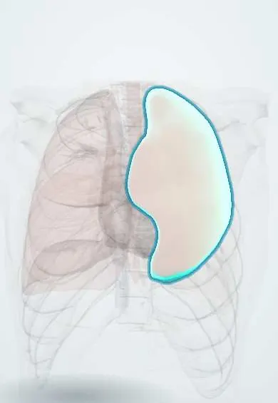
Left upper lobe !
The left upper lobe fills the upper and front left chest; its tongue-shaped lower part, the lingula, takes the place of a middle lobe.
Official name: Superior lobe of left lung
Opens in the Durgical app, no sign-up needed.
What it is
The upper lobe of the left lung, including the tongue-shaped lingula that wraps in front of the heart.
What it does
Fills the upper and front left chest.
Key facts
- Where
- Above oblique fissure; segments apicoposterior, anterior, superior and inferior lingular; cardiac notch and lingula
- Blood supply
- Pulmonary artery branches (gas exchange); bronchial arteries (nourish lung tissue)
- Nerve
- Pulmonary plexus: vagus (bronchoconstriction, secretion); upper thoracic sympathetic (bronchodilation)
- Function
- Gas exchange; lingula is the left counterpart of the right middle lobe
Relations
- Above
- Cervical pleura and suprapleural membrane, with the left subclavian artery grooving the apex
- Medial
- Aortic arch, which grooves the lobe above the hilum; left common carotid and left subclavian arteries above; left vagus and phrenic nerves crossing the arch
- In front
- The lingula curves in front of the heart; at the cardiac notch the pericardium over the right ventricle touches the chest wall
- Below
- Left lower lobe across the oblique fissure; the lingula rests on the front of the left dome of the diaphragm
How to find it
The apex is heard above the medial third of the clavicle. In front, nearly the whole left chest above about the 6th costal cartilage belongs to this lobe; behind, only the part above about T3. The lingula is heard beside the lower left sternum, around the cardiac notch, and in the axilla.
On imaging
Collapse moves the lobe forward against the front chest wall rather than straight up: a hazy veil over the left chest, sometimes with a crescent of air outlining the aortic knob (Luftsichel sign). Lingular consolidation blurs the left heart border while the left hemidiaphragm stays sharp.
Common problems
- Pneumonia (infection), asthma and COPD (narrowed airways).
- Lung cancer, often linked to smoking.
- Pulmonary embolism: a clot blocks an artery in the lung.
How it is diagnosed
Listening to the chest, chest X-ray, CT, breathing tests (spirometry), blood oxygen.
Treatment
- Depends on the cause: antibiotics, inhalers, oxygen; surgery removes a lobe or segment for some cancers.
Clinical notes
- A tumour in the aortopulmonary window, between the aortic arch and the left pulmonary artery, can catch the left recurrent laryngeal nerve and cause hoarseness
- Pancoast tumour at the left apex gives the same picture as on the right: brachial plexus pain and weakness, Horner syndrome and rib destruction
- Lingula-sparing upper division segmentectomy or lingulectomy are segment-based alternatives to removing the whole lobe
Anatomical variants
- A left horizontal (minor) fissure can separate the lingula from the rest of the lobe, mimicking a left middle lobe
Development
Around week 5 the lung buds split into lobar buds: three on the right and two on the left, which is why the left lung lacks a middle lobe. On the left, the upper bud forms this lobe, including the lingula.
Related conditions
- Tuberculosis
- Sarcoidosis
- Pneumoconioses (silicosis, asbestosis, coal workers' pneumoconiosis)
- Hypersensitivity pneumonitis
- Eosinophilic pneumonias (including Löffler syndrome)
- Chronic obstructive pulmonary disease (COPD)
- Pneumothorax
- Pulmonary tuberculosis
- Lung cancer
Test yourself
Think of the answer, then tap the question to see it.
Which part of the left upper lobe takes the place of the absent middle lobe, and which outline does its disease blur on a frontal X-ray?
The lingula; lingular consolidation blurs the left heart border while the left hemidiaphragm stays sharp.
What does the cardiac notch of the left upper lobe leave uncovered, and what clinical area does this create?
Part of the pericardium near the 4th and 5th left intercostal spaces, which touches the chest wall: the area of superficial cardiac dullness.
What is the Luftsichel sign in collapse of the left upper lobe?
A crescent of air outlining the aortic knob, as the collapsed lobe moves forward and forms a hazy veil over the left chest.
Why can a tumour in the aortopulmonary window beside the left upper lobe cause hoarseness?
It can catch the left recurrent laryngeal nerve, which hooks around the aortic arch between the arch and the left pulmonary artery.
Educational information, not medical advice. For any symptom, see a health professional.
Its names
- Official Latin (TA2)
- lobus superior pulmonis sinistri; lingula pulmonis sinistri TA2 3300
- Official Spanish (DeCS)
- Lóbulo superior del pulmón izquierdo DeCS D008168
- English (TA2)
- Superior lobe of left lung
- Also called
- upper lobe of left lung · lobus ventrocranialis
Sources: Terminologia Anatomica (TA2); DeCS; Anatomy, Thorax, Lungs; 22.7 Embryonic Development of the Respiratory System (OpenStax (Rice University), 2022) · adapted, CC BY 4.0; Anatomy of the Human Body (20th ed.): The Respiratory Apparatus (Lea & Febiger (Gray's Anatomy, 1918), 1918) · adapted, Public domain.
Spotted a mistake?We are always improving for the community. Tell us what is wrong and our team will review it.Report a mistake
Representative model, for educational purposes only.