Conditions · Ophthalmology and ENT
Herpes zoster ophthalmicus !
Shingles in the first branch of the trigeminal nerve, around the eye and forehead. It can damage the cornea and the inside of the eye, so it needs eye review.
Updated 2026-11-01
Structures involved
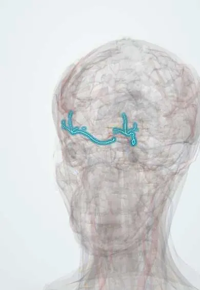
Tap one to see what it is and open it in 3D.
What it is
Reactivation of varicella-zoster virus in the trigeminal ganglion affecting the ophthalmic division (V1), with a painful vesicular rash on the forehead, upper eyelid and nose and frequent eye involvement.
Who gets it
About 10–20% of all herpes zoster involves the ophthalmic nerve. Incidence rises with age and immunosuppression; in Latin America it is an important presentation in people with HIV, in whom it may appear at a younger age and be the first sign of infection.
Causes
- Reactivation of latent varicella-zoster virus in the trigeminal (Gasserian) ganglion
- Spread along the ophthalmic nerve and its branches (frontal, lacrimal, nasociliary)
Risk factors
- Age over 50
- HIV infection and other immunosuppression (cancer, transplant, corticosteroids and immunosuppressive drugs)
- Physical or emotional stress
- No previous zoster vaccination
Pathophysiology
The virus reactivates in the ganglion and travels down V1, causing neural inflammation and dermatomal vesicles. Involvement of the nasociliary branch, which supplies both the tip of the nose and the eye, predicts ocular disease. Damage occurs through direct viral infection, immune-mediated inflammation and vasculitis, affecting the eyelids, conjunctiva, cornea, uvea, retina and optic nerve.
Classification
- Skin and eyelid only
- Anterior segment: conjunctivitis, epithelial pseudodendritic keratitis, stromal and neurotrophic keratitis, anterior uveitis with raised pressure, scleritis
- Posterior segment and neuro-ophthalmic: acute retinal necrosis, optic neuritis, cranial nerve palsies
Symptoms and signs
- Prodrome of burning or tingling pain, headache and malaise on one side of the forehead
- Unilateral vesicular rash in the V1 dermatome, not crossing the midline
- Swollen eyelid, red eye, tearing and photophobia
- Blurred vision and eye pain
- Vesicles on the tip or side of the nose (Hutchinson sign)
Diagnosis
- Clinical
- Clinical diagnosis from the dermatomal rash. Every patient needs an eye examination: acuity, pupils, slit lamp with fluorescein (pseudodendrites), corneal sensation, intraocular pressure and dilated fundoscopy.
- Tests
- PCR of vesicle fluid or tears in atypical cases. HIV testing, especially in younger adults or severe disease.
- Imaging
- Not routine; brain MRI or vascular imaging when neurological signs suggest encephalitis or arteritis.
Differential diagnosis
- Herpes simplex keratitis or skin infection
- Contact dermatitis or impetigo
- Periorbital or orbital cellulitis
- Bacterial conjunctivitis
- Trigeminal neuralgia (pain without rash)
Goal of treatment
Start antivirals early to reduce viral spread and ocular complications, control inflammation and eye pressure, relieve pain and prevent postherpetic neuralgia.
Treatment
- Medical
- Oral antivirals (acyclovir, valacyclovir or famciclovir), ideally within 72 hours of rash onset but often started later if there is eye involvement; intravenous acyclovir in immunosuppressed patients or severe disease.
- Medical
- Topical corticosteroids under ophthalmology supervision for stromal keratitis and uveitis, with cycloplegics and pressure-lowering drops when needed.
- Medical
- Pain control and treatment of postherpetic neuralgia (analgesics, tricyclic antidepressants, gabapentinoids).
- Conservative
- Skin care to prevent bacterial superinfection; lubricants and lid protection for neurotrophic cornea.
- Follow-up
- Ophthalmology follow-up, as keratitis and uveitis can recur or appear months later; HIV screening and infection-control precautions for non-immune contacts.
Complications
- Chronic or recurrent keratitis, neurotrophic keratopathy and corneal scarring
- Uveitis with secondary glaucoma; cataract
- Acute retinal necrosis and optic neuritis
- Eyelid scarring, ptosis and cranial nerve palsies
- Postherpetic neuralgia
- Zoster-associated cerebral vasculopathy and stroke
Prognosis
Most patients treated early keep good vision, but ocular inflammation can become chronic or recurrent and needs long-term follow-up. Postherpetic neuralgia is common in older adults.
Prevention
- Recombinant zoster vaccine for older adults and eligible immunocompromised people
- Childhood varicella vaccination reduces circulating virus
Test yourself
Think of the answer, then tap the question to see it.
What is Hutchinson sign in herpes zoster ophthalmicus?
Vesicles on the tip, side or root of the nose, showing nasociliary nerve involvement and predicting eye disease.
Which nerve and ganglion are involved in herpes zoster ophthalmicus?
The ophthalmic division (V1) of the trigeminal nerve, with virus reactivating in the trigeminal ganglion.
What is the first-line treatment for herpes zoster ophthalmicus?
Oral antivirals (acyclovir, valacyclovir or famciclovir), ideally started within 72 hours, plus ophthalmology review.
In a young adult with herpes zoster ophthalmicus, which infection should be tested for?
HIV infection.
Related conditions
- Herpes zoster (shingles)
- Varicella (chickenpox)
- Keratitis and corneal ulcer
- Uveitis
- HIV infection and AIDS
- Acute angle-closure glaucoma
- Ischaemic stroke
Educational information, not medical advice. For any symptom, see a health professional.
Sources: Herpes Zoster Ophthalmicus (EyeWiki (American Academy of Ophthalmology), 2025); Clinical Overview of Shingles (Herpes Zoster) (Centers for Disease Control and Prevention (CDC), 2024).
Spotted a mistake?We are always improving for the community. Tell us what is wrong and our team will review it.Report a mistake
Representative model, for educational purposes only.