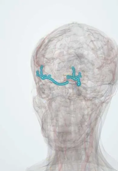
Nervous system · Head and neck
V1 ophthalmic !
The ophthalmic nerve (V1), the top branch of the trigeminal nerve, gives feeling to the forehead, upper eyelid, eye surface and nose tip.
Official name: Ophthalmic nerve
Opens in the Durgical app, no sign-up needed.
What it is
The top trigeminal branch, to the forehead, eye and nose.
What it does
Feeling of the forehead, upper lid, cornea and nose tip; shingles here threatens the eye.
Key facts
- Roots
- V1; superior orbital fissure (frontal, lacrimal, nasociliary branches)
- Moves
- None
- Feeling from
- Forehead, upper eyelid, cornea (corneal reflex), nose to the tip
Relations
- Course
- From the trigeminal ganglion forward in the lateral wall of the cavernous sinus; just before the superior orbital fissure it splits: the lacrimal and frontal nerves pass above the common tendinous ring, the nasociliary nerve through it; the frontal nerve ends on the forehead as the supraorbital and supratrochlear nerves
- Above
- In the cavernous sinus wall, the oculomotor nerve (III) and the trochlear nerve (IV)
- Below
- The V2 maxillary nerve, lowest in the lateral wall of the cavernous sinus
- Medial
- Within the cavernous sinus, the internal carotid artery and the abducens nerve (VI)
How to find it
Its skin territory runs from the upper eyelid and the bridge and tip of the nose up over the forehead to the vertex. The supraorbital notch is felt on the upper orbital margin at the junction of its inner third and outer two thirds.
Examination
- Corneal reflex: touching the edge of the cornea makes both eyes blink; no blink in either eye when one side is touched means an afferent (V1) lesion on that side, while one eye that never blinks means a facial nerve problem on that side
- Light touch and pinprick on the forehead, compared with the other side
On imaging
Seen mostly through its surroundings. Contrast MRI, coronal and axial, follows it in the lateral wall of the cavernous sinus to the superior orbital fissure; a mass or thrombosis in the sinus widens that wall or alters its enhancement. CT with bone windows shows the supraorbital notch and orbital roof fractures. In herpes zoster ophthalmicus, imaging is for complications.
Clinical notes
- Zoster ophthalmicus can cause keratitis, uveitis and corneal scarring, so the eye is examined even when only the skin seems affected
- Frontal and ethmoid sinusitis is felt over the forehead and between the eyes through V1 branches
- Superior orbital fissure syndrome: III, IV, V1 and VI affected together, with a frozen eye, ptosis and forehead numbness
Anatomical variants
- The supraorbital nerve leaves through a notch in many people and through a complete bony foramen in others; a foramen can sit a little above the orbital margin, which matters when placing a block
Memory aid
Its three branches: "NFL": Nasociliary, Frontal, Lacrimal.
Related conditions
Test yourself
Think of the answer, then tap the question to see it.
What is the Hutchinson sign in herpes zoster ophthalmicus?
Vesicles on the tip or side of the nose: the nasociliary branch of V1 is involved and the eye is at higher risk.
What are the three branches of the V1 ophthalmic nerve?
Nasociliary, frontal and lacrimal (NFL); the frontal ends as the supraorbital and supratrochlear nerves.
In the corneal reflex, what does no blink in either eye when one cornea is touched indicate?
An afferent lesion of the V1 ophthalmic nerve on that side; one eye that never blinks points to the facial nerve.
Which nerves are affected together in superior orbital fissure syndrome?
III, IV, V1 and VI: a frozen eye, ptosis and forehead numbness.
Educational information, not medical advice. For any symptom, see a health professional.
Its names
- Official Latin (TA2)
- nervus ophthalmicus TA2 6196
- Official Spanish (DeCS)
- Nervio Oftálmico DeCS D009882
- English (TA2)
- Ophthalmic nerve
- Also called
- V1 · cranial nerve V1 · ophthalmic division of trigeminal nerve
Related
Sources: Terminologia Anatomica (TA2); DeCS; Neuroanatomy, Cranial Nerve 5 (Trigeminal); Anatomy, Head and Neck: Eye Nerves; Anatomy of the Human Body (20th ed.): The Exterior of the Skull (Lea & Febiger (Gray's Anatomy, 1918), 1918) · adapted, Public domain.
Spotted a mistake?We are always improving for the community. Tell us what is wrong and our team will review it.Report a mistake
Representative model, for educational purposes only.