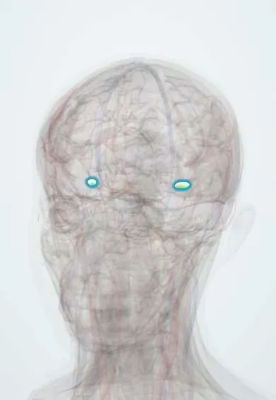
Nervous system · Head and neck
Trigeminal ganglion !
The trigeminal ganglion, at the skull base, is where the three trigeminal branches meet; the shingles virus can lie dormant here for years.
Opens in the Durgical app, no sign-up needed.
What it is
A crescent-shaped cluster of sensory nerve cells inside the skull, where the three branches of the trigeminal nerve (ophthalmic, maxillary, mandibular) come together.
What it does
Carries touch, pain and temperature from the face, the eye surface, the teeth and the inside of the mouth toward the brainstem.
Key facts
- Roots
- Sensory ganglion of the trigeminal nerve (CN V) in Meckel's cave, on the apex of the petrous temporal bone; cell bodies of the sensory fibres
- Moves
- None of its own; the motor root of V3 passes beneath it
- Feeling from
- Gives V1 (ophthalmic), V2 (maxillary) and V3 (mandibular): face, eye, nasal and oral cavities, teeth; target of procedures for trigeminal neuralgia
Relations
- Medial
- The internal carotid artery and the back of the cavernous sinus
- Below
- The motor root of the trigeminal nerve and the greater petrosal nerve, on the petrous apex of the temporal bone
- Above
- The temporal lobe, above the dural roof of Meckel's cave
- In front
- Its three divisions fan out forward: the V1 ophthalmic and V2 maxillary nerves into the lateral wall of the cavernous sinus, the V3 mandibular nerve down to the foramen ovale
- Behind
- The sensory root, running back across the petrous apex to the pons
How to find it
Deep in the middle cranial fossa and not palpable. In a middle fossa approach it is found by lifting the temporal lobe and opening the dura over the petrous apex, where the three divisions converge on it.
Examination
- Facial sensation with a cotton wisp and a pin on the forehead, cheek and chin: loss in all three divisions on one side points to the ganglion or the root rather than one branch; the skin over the angle of the jaw is spared because C2–C3 supply it
- Corneal reflex: touching one cornea normally makes both eyes blink; if neither eye blinks when one side is touched but both blink from the other side, the afferent limb (V1) on the first side is affected
On imaging
MRI shows Meckel's cave as a pocket of bright fluid on T2 around the ganglion; loss of that fluid signal or enlargement of the cave suggests a tumour (schwannoma, meningioma) or cancer spreading along a nerve.
Common problems
- Trigeminal neuralgia: brief, electric-shock-like facial pain, often triggered by touching the face or chewing.
- Shingles of the first branch can affect the forehead and the eye.
Clinical notes
- Percutaneous techniques (radiofrequency, glycerol injection, balloon compression) enter the skin a short distance lateral to the corner of the mouth and aim for the foramen ovale
- Schwannomas and meningiomas in Meckel's cave can cause facial numbness or neuralgia-like pain
- After shingles, pain can persist in the affected division once the rash has healed (postherpetic neuralgia)
Development
Forms from neural crest cells and the trigeminal placode, the ectoderm thickening that gives many of its sensory neurons.
Related conditions
Test yourself
Think of the answer, then tap the question to see it.
Where does the trigeminal ganglion lie?
In Meckel's cave, on the apex of the petrous temporal bone in the middle cranial fossa.
Which trigeminal sensory cell bodies are not in the trigeminal ganglion?
Those for proprioception from the chewing muscles: they lie in the mesencephalic nucleus in the brainstem.
Which virus lies dormant in the trigeminal ganglion, and which division is most often affected on reactivation?
Varicella-zoster virus, after chickenpox; reactivation causes shingles, most often along V1 (ophthalmic).
Through which foramen do percutaneous procedures for trigeminal neuralgia reach the trigeminal ganglion?
The foramen ovale, with a needle entering the skin a short distance lateral to the corner of the mouth.
Educational information, not medical advice. For any symptom, see a health professional.
Sources: Neuroanatomy, Semilunar Ganglion; Neuroanatomy, Cranial Nerve 5 (Trigeminal); 14.3 Motor Responses (OpenStax (Rice University), 2022) · adapted, CC BY 4.0; 16.3 The Cranial Nerve Exam (OpenStax (Rice University), 2022) · adapted, CC BY 4.0.
Spotted a mistake?We are always improving for the community. Tell us what is wrong and our team will review it.Report a mistake
Representative model, for educational purposes only.