Conditions · Hematology and oncology
Inherited thrombophilia !
Inherited gene changes that make blood clot too easily, raising the risk of deep vein thrombosis and pulmonary embolism, often at a young age.
Updated 2026-11-01
Structures involved
Tap one to see what it is and open it in 3D.
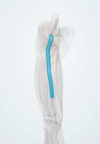
- Femoral vein
- Great saphenous vein
- Popliteal vein
- Deep femoral vein
- Small saphenous vein
- Pulmonary arteries
- Portal vein
In the clinical exam
Why it happens
- Thrombosis and Virchow's triad
- Hypercoagulable states: inherited and acquired
- Fibrinolysis and natural anticoagulants
- The coagulation cascade
What it is
A group of inherited conditions that increase the tendency to form venous blood clots, either by making procoagulant factors resistant or more abundant (factor V Leiden, prothrombin G20210A) or by reducing natural anticoagulants (antithrombin, protein C, protein S). Antiphospholipid syndrome is an acquired thrombophilia and is covered separately.
Who gets it
Factor V Leiden is the most common inherited thrombophilia in people of European ancestry (heterozygous in about 3 to 8%) and the prothrombin G20210A variant is second. Both are less frequent in Indigenous and African-descended populations; Latin American studies show intermediate frequencies reflecting mixed ancestry. Antithrombin, protein C and protein S deficiencies are rare but carry a higher risk.
Causes
- Factor V Leiden: a point mutation (R506Q) that makes factor V resistant to activated protein C
- Prothrombin G20210A: a variant that raises prothrombin levels
- Antithrombin deficiency
- Protein C deficiency
- Protein S deficiency
- Usually autosomal dominant; homozygous or combined defects carry a much higher risk
Risk factors
- Family history of venous thrombosis, especially in young relatives
- Added acquired triggers: surgery, trauma, immobility, cancer, long travel
- Oestrogen-containing contraceptives and hormone therapy
- Pregnancy and the postpartum period
- Obesity and older age
Pathophysiology
Normal clotting is balanced by natural anticoagulants. Activated protein C, with its cofactor protein S, inactivates factors Va and VIIIa; antithrombin neutralises thrombin and factor Xa. Factor V Leiden resists this inactivation (activated protein C resistance), the prothrombin variant increases thrombin generation, and deficiencies of antithrombin, protein C or S remove brakes. Clots form mainly in the deep veins of the legs, but also in unusual sites (cerebral, portal, mesenteric or hepatic veins). Protein C deficiency explains warfarin-induced skin necrosis, and antithrombin deficiency can cause resistance to heparin.
Classification
- Gain of function: factor V Leiden, prothrombin G20210A (common, milder risk)
- Loss of function: antithrombin, protein C and protein S deficiencies (rare, higher risk)
- Heterozygous versus homozygous or combined defects
- Distinguish from acquired thrombophilias: antiphospholipid syndrome, cancer, nephrotic syndrome, PNH, myeloproliferative neoplasms
Symptoms and signs
- Many carriers never have a clot
- Painful, swollen, warm leg from deep vein thrombosis
- Sudden breathlessness, chest pain or haemoptysis from pulmonary embolism
- Severe headache or seizures from cerebral venous thrombosis
- Recurrent pregnancy loss or placental complications in some women
- Purpura fulminans in newborns with homozygous protein C deficiency
Diagnosis
- Clinical
- Suspect it with venous thrombosis at a young age, a weak or absent trigger, unusual sites, recurrence or a strong family history. Testing does not change acute treatment, and routine testing after a first provoked clot is not recommended.
- Tests
- Genetic tests for factor V Leiden (or activated protein C resistance assay) and prothrombin G20210A, which are reliable at any time; functional levels of antithrombin, protein C and protein S, which are best measured away from the acute clot and off anticoagulants (heparin lowers antithrombin; warfarin lowers protein C and S; pregnancy and oestrogens lower protein S). Antiphospholipid antibodies to exclude the acquired form.
- Imaging
- Compression Doppler ultrasound for deep vein thrombosis; CT pulmonary angiography for pulmonary embolism; brain MR venography or CT venography for cerebral venous thrombosis.
Differential diagnosis
- Antiphospholipid syndrome
- Cancer-associated thrombosis
- Myeloproliferative neoplasms (polycythaemia vera, essential thrombocythaemia)
- Paroxysmal nocturnal haemoglobinuria
- Heparin-induced thrombocytopenia
- Nephrotic syndrome and oestrogen-related thrombosis
Goal of treatment
Treat each clot with standard anticoagulation, decide the duration by weighing recurrence against bleeding risk, and prevent clots in high-risk situations.
Treatment
- Medical
- Acute venous thrombosis is treated as in anyone else: parenteral anticoagulation (low molecular weight heparin) followed by an oral anticoagulant (a direct oral anticoagulant or warfarin overlapped with heparin).
- Medical
- Long-term or indefinite anticoagulation is considered after unprovoked or recurrent clots and in high-risk defects (antithrombin deficiency, homozygous or combined defects); antithrombin concentrate in selected situations.
- Conservative
- Thromboprophylaxis during surgery, immobility, pregnancy and the postpartum period in affected carriers according to risk; avoid oestrogen-containing contraceptives in favour of non-oestrogen methods.
- Lifestyle
- Keep active, stay hydrated and move the legs on long trips, maintain a healthy weight and stop smoking.
- Follow-up
- Nursing priorities: monitor for bleeding on anticoagulation, INR when warfarin is used, platelet count on heparin, and teach recognition of leg and chest symptoms; check skin after starting warfarin in protein C deficiency.
Complications
- Recurrent deep vein thrombosis
- Pulmonary embolism and chronic thromboembolic pulmonary hypertension
- Post-thrombotic syndrome
- Pregnancy complications (pregnancy loss, preeclampsia, placental abruption) in some women
- Warfarin-induced skin necrosis
- Bleeding from long-term anticoagulation
Prognosis
Most heterozygous carriers of factor V Leiden or the prothrombin variant never develop a clot. Risk rises sharply with combined defects, homozygosity, antithrombin deficiency and added triggers such as pregnancy or oestrogen.
Prevention
- Prophylaxis in high-risk situations, informed choice of contraception, and family counselling
- Screening of relatives only when the result would change management
Test yourself
Think of the answer, then tap the question to see it.
What is the most common inherited thrombophilia in people of European ancestry?
Factor V Leiden, which causes resistance to activated protein C.
Which deficiency causes resistance to heparin?
Antithrombin deficiency, because heparin works by enhancing antithrombin.
Which deficiency predisposes to skin necrosis when warfarin is started without heparin overlap?
Protein C deficiency.
Why are protein C, protein S and antithrombin levels not measured during acute thrombosis and anticoagulation?
The clot and the anticoagulants lower their levels, giving falsely low results.
Which contraceptive type should carriers of inherited thrombophilia avoid?
Combined oestrogen-containing contraceptives.
Related conditions
- Deep vein thrombosis
- Pulmonary embolism
- Cerebral venous sinus thrombosis
- Portal vein thrombosis
- Budd-Chiari syndrome
- Miscarriage (early pregnancy loss)
- Preeclampsia
- Heparin-induced thrombocytopenia
Educational information, not medical advice. For any symptom, see a health professional.
Sources: Factor V Leiden thrombophilia (MedlinePlus (U.S. National Library of Medicine), 2023); Prothrombin thrombophilia (MedlinePlus (U.S. National Library of Medicine), 2025).
Spotted a mistake?We are always improving for the community. Tell us what is wrong and our team will review it.Report a mistake
Representative model, for educational purposes only.