Conditions · Gastroenterology and hepatology
Microscopic colitis !
Inflammation of the colon that looks normal on colonoscopy but shows changes under the microscope. It causes chronic watery diarrhoea, often in older women.
Updated 2026-11-01
Structures involved
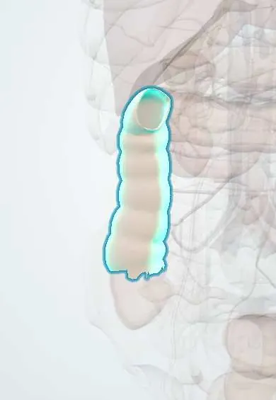
Tap one to see what it is and open it in 3D.
Why it happens
- Chronic inflammation: causes, cells and features
- Water and electrolyte absorption and how diarrhea happens
- Large intestine, appendix and anal canal
What it is
A chronic inflammatory disease of the colon that causes watery, non-bloody diarrhoea. The lining looks normal or nearly normal at colonoscopy, so the diagnosis depends on biopsies that show inflammation of the mucosa.
Who gets it
A common cause of chronic watery diarrhoea in older adults, usually diagnosed after age 50 and more often in women. Recognition has increased in Europe and North America; data from Latin America are limited, and it is probably under-diagnosed where colonoscopy is done without random biopsies.
Causes
- Abnormal immune response of the colonic mucosa to luminal factors in a genetically susceptible person
- Drug-associated cases: NSAIDs, proton pump inhibitors, SSRIs, statins and some other drugs are linked
- Associated autoimmune diseases (celiac disease, thyroid disease, rheumatoid arthritis, type 1 diabetes)
- Bile acid malabsorption contributes in some patients
Risk factors
- Age over 50
- Female sex (especially collagenous colitis)
- Smoking
- Use of NSAIDs, proton pump inhibitors or SSRIs
- Celiac disease or other autoimmune disease
Pathophysiology
Immune activation in the colonic mucosa increases lymphocytes in the epithelium and lamina propria. In the collagenous type, a thickened band of collagen forms under the surface epithelium. Inflammation reduces sodium and water absorption and increases chloride secretion, so the colon cannot dry the stool, producing high-volume watery diarrhoea without ulcers or bleeding.
Classification
- Collagenous colitis: thickened subepithelial collagen band (over about 10 micrometres)
- Lymphocytic colitis: increased intraepithelial lymphocytes (over 20 per 100 epithelial cells) without a thick collagen band
- Incomplete microscopic colitis: milder changes that do not meet full criteria
Symptoms and signs
- Chronic or relapsing watery, non-bloody diarrhoea, often several times a day
- Urgency, faecal incontinence and nocturnal stools
- Crampy abdominal pain and bloating
- Mild weight loss and fatigue
- Joint pains in some patients
Diagnosis
- Clinical
- Suspected in an older adult, usually a woman, with chronic watery diarrhoea, often after starting a new drug. Infection and other causes are excluded first.
- Tests
- Stool tests for infection including Clostridioides difficile, celiac serology, thyroid function and blood count. The key test is colonoscopy with multiple random biopsies from the right and left colon, even when the mucosa looks normal; histology confirms and subtypes the disease. Faecal calprotectin may be mildly raised but is not diagnostic.
- Imaging
- Colonoscopy typically shows normal or almost normal mucosa (occasionally subtle oedema or mucosal tears). Cross-sectional imaging is not needed unless another cause is suspected.
Differential diagnosis
- Irritable bowel syndrome with diarrhoea
- Celiac disease
- Infectious diarrhoea (including Clostridioides difficile and Giardia)
- Bile acid diarrhoea
- Ulcerative colitis or Crohn disease
- Lactose intolerance and laxative use
Goal of treatment
Achieve clinical remission (fewer, formed stools), remove triggers and prevent relapse with the lowest effective treatment.
Treatment
- Conservative
- Stop or replace suspected drugs (NSAIDs, proton pump inhibitors, SSRIs) and stop smoking; reduce caffeine, alcohol and lactose if they worsen symptoms.
- Medical
- Oral budesonide, a gut-targeted corticosteroid, is first-line for induction and, if relapses occur, maintenance at the lowest effective level.
- Medical
- Antidiarrhoeal agents (loperamide), bismuth salts or bile acid sequestrants for mild disease or as add-on; immunomodulators or biological therapy for rare refractory cases.
- Follow-up
- Track stool frequency, hydration, potassium and weight; screen for celiac disease; ensure fall prevention and skin care in older patients with urgency and incontinence.
Complications
- Dehydration and electrolyte disturbances (low potassium)
- Faecal incontinence and reduced quality of life
- Frequent relapse after stopping treatment
- Corticosteroid side effects with long-term use (bone loss)
Prognosis
Benign: it does not raise the risk of colorectal cancer and does not cause strictures. Most patients respond to treatment, but relapse is common and some need long-term maintenance.
Prevention
- Avoiding unnecessary long-term NSAIDs and proton pump inhibitors
- Not smoking
Test yourself
Think of the answer, then tap the question to see it.
What does colonoscopy usually show in microscopic colitis?
Normal or nearly normal mucosa; the diagnosis needs random biopsies.
What are the two histological subtypes?
Collagenous colitis (thick subepithelial collagen band) and lymphocytic colitis (increased intraepithelial lymphocytes).
What is the typical patient?
An older woman with chronic watery, non-bloody diarrhoea, often taking NSAIDs, proton pump inhibitors or SSRIs.
What is first-line drug treatment?
Oral budesonide.
Related conditions
- Coeliac disease
- Irritable bowel syndrome
- Crohn disease
- Ulcerative colitis
- Clostridioides difficile infection
- Lactose intolerance
Educational information, not medical advice. For any symptom, see a health professional.
Sources: Microscopic Colitis - NIDDK (National Institute of Diabetes and Digestive and Kidney Diseases (NIDDK), 2025).
Spotted a mistake?We are always improving for the community. Tell us what is wrong and our team will review it.Report a mistake
Representative model, for educational purposes only.