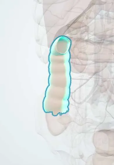
Ascending colon !
The ascending colon is the part of the large intestine that runs up the right side of the belly, from the cecum to just under the liver.
Opens in the Durgical app, no sign-up needed.
What it is
The part of the colon rising up the right side from the cecum to under the liver.
What it does
Absorbs water from liquid contents.
Key facts
- Where
- Right flank, from cecum to right colic (hepatic) flexure; secondarily retroperitoneal; right paracolic gutter lateral to it
- Blood supply
- Ileocolic and right colic arteries (SMA); marginal artery
- Nerve
- Superior mesenteric plexus (sympathetic) and vagus (parasympathetic)
- Function
- Absorbs water and electrolytes; moves liquid contents upward
Relations
- In front
- Coils of ileum, greater omentum and the anterior abdominal wall
- Behind
- Iliacus, quadratus lumborum and the lower pole of the right kidney, with the iliohypogastric, ilioinguinal and lateral femoral cutaneous nerves
- Medial
- Loops of ileum; deeper, the right ureter and gonadal vessels
- Lateral
- Right paracolic gutter and the white line of Toldt
- Above
- Right colic (hepatic) flexure under the right lobe of the liver and the gallbladder, in front of the right kidney, with D2 just medial
How to find it
It projects onto the right flank. Not felt in health; a mass here in an older adult with anemia raises suspicion of right colon cancer. At surgery the lateral peritoneal reflection (white line of Toldt) is opened to lift the colon and its mesocolon off the retroperitoneum (renal fascia, duodenum, ureter).
On imaging
On CT it sits in the right flank with the right paracolic gutter lateral to it, where free fluid collects. Barium enema and CT colonography outline its haustra; an apple-core stricture suggests carcinoma.
Common problems
- Colon polyps and colorectal cancer: screening finds them early.
- Diverticular disease and diverticulitis (usually sigmoid).
- Ulcerative colitis and Crohn disease.
How it is diagnosed
Colonoscopy; CT; stool tests for screening.
Treatment
- Polyps removed during colonoscopy; surgery (colectomy) for cancer or complicated disease.
Clinical notes
- Pain from the ascending colon (midgut) is referred to the periumbilical region
- A perforated duodenal ulcer can leak down the right paracolic gutter and mimic appendicitis (Valentino syndrome)
- The bloodless fusion plane between its mesocolon and the retroperitoneum is the plane followed in a right colectomy
Development
It forms from the caudal limb of the midgut loop, beyond the cecal bud, and is supplied by the superior mesenteric artery. After the gut rotates, the cecum moves down from under the liver and the ascending mesocolon usually fuses with the back wall; when that fusion is incomplete the right colon stays mobile and can twist (cecal volvulus).
Related conditions
- Colorectal cancer
- Large bowel obstruction
- Crohn disease
- Intussusception (children)
- Lynch syndrome (hereditary non-polyposis colorectal cancer)
- Microscopic colitis
- Intestinal amoebiasis (amoebic dysentery)
- Necrotising enterocolitis
Test yourself
Think of the answer, then tap the question to see it.
How is the ascending colon mobilized in a right colectomy?
By incising the white line of Toldt in the right paracolic gutter and sweeping the colon and mesocolon medially along the bloodless fusion plane.
Which structures behind the ascending colon must be protected in a right hemicolectomy?
The second and third parts of the duodenum and the right ureter (with the gonadal vessels).
What is Valentino syndrome?
A perforated duodenal ulcer leaking down the right paracolic gutter beside the ascending colon, mimicking appendicitis.
Educational information, not medical advice. For any symptom, see a health professional.
Its names
- Official Latin (TA2)
- colon ascendens TA2 2982
- Official Spanish (DeCS)
- Colon ascendente DeCS D044682
- English (TA2)
- Ascending colon
- Also called
- right colon
Related
Sources: Terminologia Anatomica (TA2); DeCS; Anatomy, Abdomen and Pelvis: Large Intestine; Anatomy of the Human Body (20th ed.): The Digestive Apparatus (Lea & Febiger (Gray's Anatomy, 1918), 1918) · adapted, Public domain.
Spotted a mistake?We are always improving for the community. Tell us what is wrong and our team will review it.Report a mistake
Representative model, for educational purposes only.