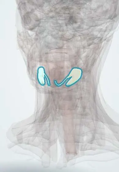
Sialolithiasis (salivary stones) !
Sialolithiasis is a stone in a salivary gland or its duct; it blocks saliva and makes the gland swell and hurt at meals, most often under the jaw.
Updated 2026-10-29
Structures involved
Tap one to see what it is and open it in 3D.
In histology
In physiology
What it is
Formation of a calculus (stone) inside a salivary gland or its duct, which blocks saliva flow and causes painful swelling of the gland at meals. It most often affects the submandibular gland.
Who gets it
The most common disease of the major salivary glands in middle-aged adults; uncommon in children.
Causes
- Calcium salts precipitating around a nucleus of mucus, debris or bacteria
- Salivary stasis
Risk factors
- Dehydration and reduced saliva flow
- Medicines that reduce saliva (anticholinergics, diuretics)
- Chronic sialadenitis
- Smoking
- Gout
Pathophysiology
Submandibular saliva is more alkaline, richer in calcium and mucin, and flows uphill through a long Wharton duct, which favours stones there. A stone blocks the duct, so saliva backs up during meals and stretches the gland. Stasis invites retrograde bacterial infection (sialadenitis).
Symptoms and signs
- Painful swelling of the gland during or just after meals (salivary colic)
- Swelling that settles over hours
- A palpable hard stone in the floor of the mouth
- Pus at the duct opening when infected
- Bad taste or dry mouth
Diagnosis
- Clinical
- History of meal-time swelling and bimanual palpation of the floor of the mouth and gland.
- Tests
- Culture of pus from the duct if infected.
- Imaging
- Ultrasound is first-line; non-contrast CT detects small stones; sialendoscopy is both diagnostic and therapeutic.
Differential diagnosis
- Acute bacterial sialadenitis
- Mumps
- Salivary gland tumour
- Cervical lymphadenitis
- Ranula
Goal of treatment
Relieve obstruction and pain, treat infection and preserve gland function.
Treatment
- Conservative
- Hydration, warm compresses, gland massage and sialogogues (such as sour sweets) to help small stones pass.
- Medical
- Analgesics; antibiotics for superimposed bacterial sialadenitis.
- Procedure
- Sialendoscopy with stone retrieval or transoral removal of stones near the duct opening.
- Surgical
- Excision of the gland for recurrent disease or deep stones that cannot be removed otherwise.
Complications
- Acute or chronic sialadenitis
- Gland abscess
- Duct stricture
- Nerve injury after gland excision (marginal mandibular, lingual or hypoglossal)
Prognosis
Most patients do well with gland-preserving treatment; stones can recur.
Prevention
- Good hydration and oral hygiene
- Review of medicines that dry the mouth
Test yourself
Think of the answer, then tap the question to see it.
Which salivary gland is most often affected by stones?
The submandibular gland, through Wharton duct.
Why do symptoms appear at meal times?
Saliva production rises with eating and backs up behind the stone, stretching the gland.
What is the first-line imaging for sialolithiasis?
Ultrasound; non-contrast CT is used for small or uncertain stones.
Which minimally invasive technique allows removal of salivary stones?
Sialendoscopy.
Related conditions
Educational information, not medical advice. For any symptom, see a health professional.
Sources: Sialolithiasis (StatPearls (NCBI Bookshelf), 2024).
Spotted a mistake?We are always improving for the community. Tell us what is wrong and our team will review it.Report a mistake
Representative model, for educational purposes only.