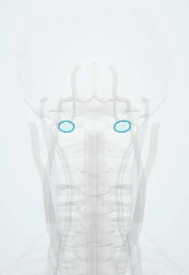
Skull–atlas joints !
The atlanto-occipital joints are the two joints between the skull and the atlas (C1); they let the head nod "yes".
Official name: Atlanto-occipital joint
Opens in the Durgical app, no sign-up needed.
What it is
The two joints between the skull and the atlas.
What it does
Allow nodding 'yes' (about 25° of bending forward and back) and very little turning.
Key facts
- Where
- Occipital condyles on the concave superior facets of the atlas lateral masses (C0–C1)
- Attaches
- Loose fibrous capsules; anterior and posterior atlanto-occipital membranes
- Function
- Synovial condyloid (ellipsoid) joints; nodding flexion-extension (about 25°) and slight side bending, almost no rotation
Relations
- Above
- Occipital condyles of the occipital bone on each side of the foramen magnum; the hypoglossal canal runs just above each condyle
- Medial
- Foramen magnum with the junction of the medulla and spinal cord, the vertebral arteries entering the skull and the alar ligaments
- In front
- Anterior atlanto-occipital membrane, rectus capitis anterior and longus capitis; in front of them, the retropharyngeal space and the pharynx
- Behind
- Posterior atlanto-occipital membrane and the suboccipital triangle (rectus capitis posterior major, obliquus capitis superior and inferior) with the vertebral artery in its groove
- Below
- Lateral masses of the atlas (C1), whose lower facets form the lateral atlantoaxial joints
On imaging
CT with sagittal and coronal reformats shows the condyle–C1 gap; widening or asymmetry suggests craniocervical dissociation. On a lateral view or CT, the basion–dens interval and the Powers ratio screen for dislocation (a Powers ratio above 1 suggests anterior dislocation). Condyle fractures are easily missed on radiographs and are best seen on CT.
Common problems
- Neck pain and headaches from arthritis or after whiplash.
- Bone spurs that narrow the opening for a nerve root.
How it is diagnosed
There is no single sign. CT or MRI show arthritis; a numbing injection of the nerves to the joint (medial branch block) shows whether the joint is the pain source.
Treatment
- Physical therapy, medicine, injections; radiofrequency ablation for lasting facet pain.
Clinical notes
- Survivors of atlanto-occipital dislocation may have brainstem, lower cranial nerve or vertebral artery injury
- Occipital condyle fractures (Anderson–Montesano types I–III) enter this joint; they are found on CT rather than plain films
- In rheumatoid arthritis, erosion of these joints and the atlantoaxial joints lets the skull settle onto C1–C2, so the dens rises toward the foramen magnum (cranial settling)
Anatomical variants
- Occipitalisation (assimilation) of the atlas: partial or complete fusion of C1 with the occipital bone; uncommon, reported frequencies vary, and it may come with basilar invagination or a Chiari malformation, adding load to C1–C2
Development
The condyles and the region around the foramen magnum come from the lowest occipital sclerotomes (the proatlas); faulty segmentation there explains atlas assimilation and a small extra bony knob in front of the foramen magnum (third condyle).
Test yourself
Think of the answer, then tap the question to see it.
What type of joint are the atlanto-occipital joints, and what movement do they allow?
Synovial condyloid (ellipsoid) joints between the occipital condyles and the atlas; they allow nodding flexion-extension (about 25°), with almost no rotation.
What mainly stabilises the atlanto-occipital joints?
Ligaments joining the skull to C2, the tectorial membrane and the alar ligaments, rather than the loose joint capsules.
Why are children more prone to atlanto-occipital dislocation?
Their head is large and their occipital condyles are flatter and smaller; the injury is high-energy and often fatal.
On imaging, what does a Powers ratio above 1 suggest?
Anterior atlanto-occipital dislocation; CT also shows widening or asymmetry of the condyle–C1 gap in craniocervical dissociation.
Educational information, not medical advice. For any symptom, see a health professional.
Its names
- Official Latin (TA2)
- articulatio atlantooccipitalis TA2 1630
- Official Spanish (DeCS)
- Articulación atlantooccipital DeCS D001269
- English (TA2)
- Atlanto-occipital joint
- Also called
- articulus atlantooccipitalis
Related
Sources: Terminologia Anatomica (TA2); DeCS; Anatomy, Head and Neck: Atlantoaxial Joint; Anatomy, Head and Neck: Foramen Magnum.
Spotted a mistake?We are always improving for the community. Tell us what is wrong and our team will review it.Report a mistake
Representative model, for educational purposes only.