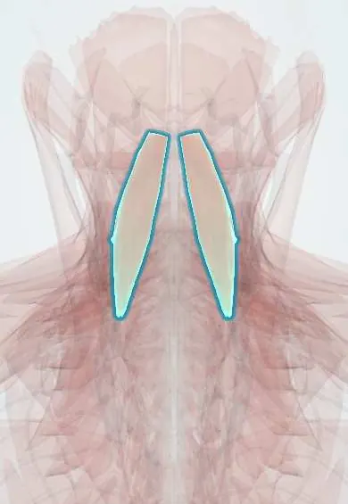
Longus capitis !
The longus capitis is a deep muscle in front of the upper neck vertebrae, running up to the skull base; it bends the head forward.
Official name: Longus capitis muscle
Opens in the Durgical app, no sign-up needed.
What it is
A deep neck muscle in front of the upper neck vertebrae, running up to the skull base.
What it does
Bends the head and upper neck forward.
Key facts
- Origin
- Anterior tubercles of transverse processes of C3–C6
- Insertion
- Basilar part of the occipital bone
- Action
- Flexes the head and upper neck
- Nerve
- Ventral rami of C1–C3
- Blood supply
- Ascending pharyngeal, ascending cervical and vertebral arteries
Relations
- In front
- Prevertebral fascia, then the retropharyngeal space and the pharynx; the sympathetic trunk with the superior cervical ganglion lies on it
- Behind
- Transverse processes of C3–C6, the atlas, rectus capitis anterior and the anterior atlanto-occipital membrane
- Medial
- Longus colli below; the anterior longitudinal ligament in the midline
- Lateral
- Carotid sheath (internal carotid artery, internal jugular vein, vagus nerve) in front and to the side; scalenus anterior below
How to find it
It cannot be felt. On sagittal and axial MRI it is the muscle band in front of C1–C3 behind the pharynx; on a lateral neck radiograph it is part of the thin prevertebral soft tissue in front of C2.
Examination
- Craniocervical flexion test: a gentle nod lying on the back, in stages, often with a pressure sensor under the neck; failing to hold the nod or substituting with sternocleidomastoid shows weak deep neck flexors
On imaging
On a lateral neck radiograph it is part of the prevertebral soft tissue in front of C2, normally thin (about 7 mm in adults is a common cut-off). Widening suggests a retropharyngeal abscess, haematoma or post-traumatic swelling; CT or MRI then shows the collection. CT also shows calcific tendinitis of the nearby longus colli: calcium in front of C1–C2 with fluid, an abscess mimic.
Common problems
- Strained in whiplash.
- In an ACDF it is gently lifted off the front of the spine on each side to place the retractor; the sympathetic trunk lies on it.
How it is diagnosed
Exam; MRI if needed.
Treatment
- Deep neck flexor exercises are a mainstay of neck pain rehabilitation.
Clinical notes
- A retropharyngeal abscess or haematoma lies just in front of it and widens the prevertebral soft tissue on the lateral radiograph
- High anterior and transoral approaches to C1–C2 cross it; the superior cervical ganglion on its surface is at risk
Test yourself
Think of the answer, then tap the question to see it.
What are the attachments and action of the longus capitis?
From the anterior tubercles of C3–C6 to the basilar occipital bone; it flexes the head and upper neck.
Which ganglion lies on the longus capitis at about C2–C3?
The superior cervical sympathetic ganglion; injury causes Horner syndrome (ptosis, miosis, anhidrosis).
Which test assesses the longus capitis and longus colli as deep neck flexors?
The craniocervical flexion test: a staged gentle nod lying supine; failing it or substituting with sternocleidomastoid shows weakness.
Educational information, not medical advice. For any symptom, see a health professional.
Its names
- Official Latin (TA2)
- musculus longus capitis TA2 2149
- Official Spanish (DeCS)
- Músculo largo de la cabeza DeCS D009334
- English (TA2)
- Longus capitis muscle
Related
Sources: Terminologia Anatomica (TA2); DeCS; Anatomy, Head and Neck, Prevertebral Muscles.
Spotted a mistake?We are always improving for the community. Tell us what is wrong and our team will review it.Report a mistake
Representative model, for educational purposes only.