Otitis externa !
Otitis externa, or swimmer's ear, is an infection of the ear canal, usually after moisture gets trapped; it causes ear pain and itching.
Updated 2026-10-29
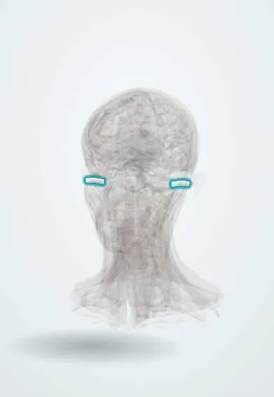
Structures involved
Tap one to see what it is and open it in 3D.
Microorganisms that cause it
In the clinical exam
What it is
Inflammation and infection of the external ear canal, usually bacterial. Often called «swimmer's ear» because moisture in the canal is the classic trigger.
Who gets it
Common at all ages and most frequent in hot, humid climates and in swimmers. Necrotising (malignant) otitis externa is a rare, serious form seen mainly in older adults with diabetes or immunosuppression.
Causes
- Pseudomonas aeruginosa (most common)
- Staphylococcus aureus
- Mixed and other gram-negative bacteria
- Fungi (Aspergillus, Candida), especially after long courses of antibiotic drops
Risk factors
- Swimming, humidity and sweating that keep the canal wet
- Trauma from cotton swabs, fingernails or cleaning
- Hearing aids and earphones
- Loss of protective cerumen
- Skin conditions of the canal (eczema, seborrhoeic dermatitis, psoriasis)
- Diabetes or immunosuppression (risk of the necrotising form)
Pathophysiology
Water and trauma break down the acidic, waxy barrier of the canal skin. Bacteria colonise the macerated skin, which swells and narrows the canal; inflammation can spread to the auricle and surrounding soft tissue. In the necrotising form, infection invades the temporal bone and skull base.
Classification
- Acute diffuse (most cases)
- Acute localised (furuncle of the canal)
- Chronic (lasting more than about 3 months)
- Otomycosis (fungal)
- Necrotising (malignant) otitis externa
Symptoms and signs
- Ear pain, often intense, worse when moving the tragus or pulling the auricle
- Itching
- Discharge from the canal
- Feeling of fullness and muffled hearing
- Swollen, red canal with debris
Diagnosis
- Clinical
- Based on history and otoscopy: tenderness on tragal pressure or pulling the auricle, oedematous red canal with debris. The eardrum, when seen, is usually normal; fungal hyphae or spores suggest otomycosis.
- Tests
- Culture of the discharge in severe, recurrent or refractory cases. Glucose and inflammatory markers when necrotising infection is suspected.
- Imaging
- Not needed in uncomplicated cases. CT and/or MRI of the temporal bone, and sometimes nuclear scans, when necrotising otitis externa is suspected.
Differential diagnosis
- Acute otitis media with perforation and discharge
- Furuncle of the ear canal
- Contact dermatitis of the canal
- Herpes zoster oticus (Ramsay Hunt syndrome)
- Cholesteatoma
- Foreign body
Goal of treatment
Clear the infection, relieve pain and restore a dry, healthy canal, while spotting the necrotising form early.
Treatment
- Medical
- Topical antibiotic drops, often with a corticosteroid, are first line; non-ototoxic drops (such as fluoroquinolones) are preferred when the eardrum may be perforated. Antifungal drops for otomycosis.
- Procedure
- Aural toilet (gentle cleaning by microsuction) so the drops reach the skin; a wick when the canal is too swollen.
- Conservative
- Keeping the ear dry, avoiding swabs and earphones during treatment, and analgesics for pain.
- Medical
- Systemic antibiotics when infection spreads beyond the canal or the patient is diabetic or immunosuppressed; prolonged antipseudomonal therapy for necrotising otitis externa.
Drugs for this condition
Complications
- Spreading cellulitis of the auricle (perichondritis)
- Chronic otitis externa and canal stenosis
- Necrotising otitis externa with skull base osteomyelitis
- Cranial nerve palsies (facial nerve first)
Prognosis
Most acute cases improve within days of correct topical treatment. Necrotising otitis externa needs long treatment and can be fatal if diagnosed late.
Prevention
- Drying the ears after swimming; earplugs for frequent swimmers
- Not using cotton swabs in the canal
- Acidifying drops in some recurrent cases
Test yourself
Think of the answer, then tap the question to see it.
Which bacterium causes most cases of acute otitis externa?
Pseudomonas aeruginosa, followed by Staphylococcus aureus.
Which examination sign helps separate otitis externa from otitis media?
Pain on pressing the tragus or pulling the auricle.
Which patient is at risk of necrotising otitis externa?
An older adult with diabetes or immunosuppression; it spreads to the temporal bone and skull base.
What is the first-line treatment of uncomplicated acute otitis externa?
Topical antibiotic drops (often with a corticosteroid) after cleaning the canal; systemic antibiotics are not routine.
Related conditions
Educational information, not medical advice. For any symptom, see a health professional.
Sources: Otitis Externa (StatPearls (NCBI Bookshelf), 2023).
Spotted a mistake?We are always improving for the community. Tell us what is wrong and our team will review it.Report a mistake
Representative model, for educational purposes only.