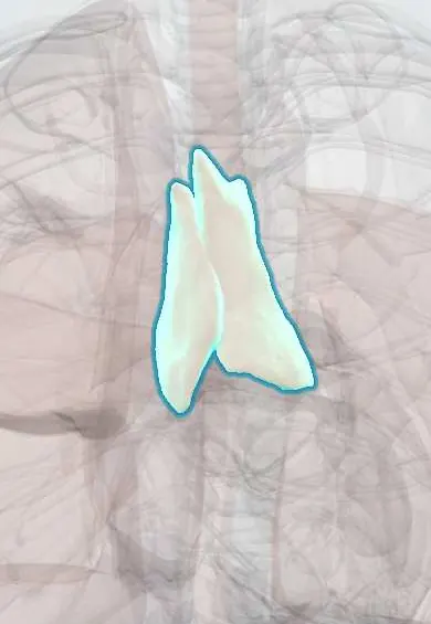
Thymus !
The thymus is a gland behind the upper breastbone that trains white blood cells (T cells) in childhood, then shrinks and is replaced by fat.
Opens in the Durgical app, no sign-up needed.
What it is
A soft gland in the upper front chest, behind the sternum; large in children, mostly replaced by fat in adults.
What it does
Trains white blood cells (T cells) to recognize the body's own tissues during childhood.
Key facts
- Where
- Superior and anterior mediastinum, behind manubrium; may extend into lower neck
- Blood supply
- Branches of internal thoracic arteries (and inferior thyroid arteries)
- Nerve
- Vagus and sympathetic branches (mainly vasomotor)
- Function
- T-lymphocyte maturation; large in childhood, replaced by fat after puberty
Relations
- In front
- Manubrium and upper body of the sternum, the sternohyoid and sternothyroid muscles and the upper costal cartilages
- Behind
- Pericardium, ascending aorta and aortic arch, the left brachiocephalic vein and, higher up, the trachea
- Lateral
- Mediastinal pleura with the phrenic nerves running along its sides
- Above
- Its upper poles may reach the lower neck in front of the trachea, toward the thyroid gland
How to find it
Not palpable. It lies behind the manubrium in the anterior mediastinum, in front of the great vessels and the upper pericardium. Surgeons reach it through a median sternotomy or by thoracoscopy, tracing its upper horns toward the thyroid and keeping clear of both phrenic nerves at its sides.
On imaging
In infants, a smooth soft-tissue shadow widening the upper mediastinum (sail sign), with wavy edges from the costal cartilages. In adults, a triangle of fat in front of the aortic arch on CT; a solid nodule there points to thymoma or another anterior mass, and MRI chemical-shift imaging helps tell hyperplasia from tumour.
Common problems
- Thymoma, a usually slow-growing tumour, sometimes linked to myasthenia gravis.
How it is diagnosed
CT or MRI.
Treatment
- Surgical removal (thymectomy).
Clinical notes
- Thymectomy improves myasthenia gravis in many patients, even without a thymoma
- Removed through a median sternotomy or by thoracoscopic or robotic surgery; the phrenic nerves on both sides are the structures to protect
- Rebound thymic hyperplasia after chemotherapy or severe illness can mimic a tumour on imaging
- Thymoma is linked to other autoimmune syndromes, such as pure red cell aplasia and low antibody levels (Good syndrome)
Anatomical variants
- Extension into the neck toward the thyroid gland, seen on ultrasound in about a third of children
- Ectopic thymic tissue along its path of descent, from the angle of the mandible to the mediastinum
Development
Arises from the endoderm of the 3rd pharyngeal pouches, together with the inferior parathyroid glands, and descends into the chest; in DiGeorge syndrome (22q11 deletion) it fails to develop, with T-cell deficiency and low calcium.
Memory aid
The 4 Ts of an anterior mediastinal mass: Thymoma, Teratoma, Thyroid and Terrible lymphoma.
Related conditions
- Adenosine deaminase deficiency (ADA-SCID)
- Pure red cell aplasia (incl. Diamond–Blackfan anaemia)
- Myasthenia gravis
- DiGeorge syndrome (22q11.2 deletion syndrome)
- Mediastinal masses (thymoma and anterior mediastinal mass)
- Tetralogy of Fallot
Test yourself
Think of the answer, then tap the question to see it.
In which mediastinal compartment does the thymus lie, and which masses present there?
The anterior mediastinum (behind the manubrium); thymoma, teratoma, retrosternal thyroid and lymphoma present there as masses.
Which autoimmune disease is linked to thymoma?
Myasthenia gravis: about a third to a half of people with a thymoma have it, and thymectomy improves it in many patients.
Which nerves must be protected during thymectomy?
Both phrenic nerves, which run along the sides of the thymus on the mediastinal pleura.
From which embryonic structure does the thymus arise, and what happens in DiGeorge syndrome?
From the 3rd pharyngeal pouches, with the inferior parathyroids; in DiGeorge syndrome (22q11 deletion) it fails to develop, giving T-cell deficiency and low calcium.
What is the thymic sail sign on an infant's chest X-ray?
The large normal infant thymus widening the upper mediastinum like a sail, which can be mistaken for a mass or pneumonia.
Educational information, not medical advice. For any symptom, see a health professional.
Its names
- Official Latin (TA2)
- thymus TA2 5152
- Official Spanish (DeCS)
- Timo DeCS D013950
- English (TA2)
- Thymus
- Also called
- thymus gland
Related
Sources: Terminologia Anatomica (TA2); DeCS; Anatomy, Head and Neck, Thymus; 21.1 Anatomy of the Lymphatic and Immune Systems (OpenStax (Rice University), 2022) · adapted, CC BY 4.0.
Spotted a mistake?We are always improving for the community. Tell us what is wrong and our team will review it.Report a mistake
Representative model, for educational purposes only.