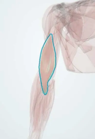
Brachialis !
The brachialis lies under the biceps at the front of the lower arm; it is the main elbow bender, whatever the position of the forearm.
Opens in the Durgical app, no sign-up needed.
What it is
A broad muscle under the biceps, from the humerus to the ulna.
What it does
The main elbow bender in every forearm position.
Key facts
- Origin
- Distal half of the anterior humerus; medial and lateral intermuscular septa
- Insertion
- Coronoid process and ulnar tuberosity
- Action
- Flexes the elbow in every forearm position
- Nerve
- Musculocutaneous nerve (C5–C6; some texts: C6–C7); small lateral part radial nerve (C7)
- Blood supply
- Brachial artery, radial recurrent artery
Relations
- Superficial
- Biceps brachii, with the musculocutaneous nerve between them; the nerve becomes the lateral forearm skin nerve at the lateral edge of the biceps tendon
- Deep
- Front of the lower humerus and the elbow capsule, to which some fibres attach
- Lateral
- Brachioradialis and ECRL, with the radial nerve and the radial recurrent artery between them
- Medial
- Brachial artery and median nerve on its surface; the medial intermuscular septum and pronator teres
- Below
- The cubital fossa, whose floor it forms, and the coronoid process
How to find it
Felt on either side of the biceps in the lower arm, best laterally, when the elbow is bent against resistance with the forearm turned palm down (which takes much of the biceps out of the movement).
Examination
- Resisted elbow flexion with the forearm pronated: weakness points to the musculocutaneous nerve or C5–C6; strength kept in mid-position suggests the brachioradialis (radial nerve) is working
On imaging
On a lateral elbow radiograph the anterior fat pad lies between it and the joint; a raised fat pad (sail sign) means an effusion. Weeks after an elbow injury, a bony mass inside the muscle in front of the distal humerus, maturing from the edge inwards, is myositis ossificans. Ultrasound or MRI show tears and haematoma before calcium appears.
Clinical notes
- In extension-type supracondylar fractures the proximal fragment can spike through the brachialis toward the skin of the fossa (pucker sign); the muscle partly shields the brachial artery and median nerve
- The anterolateral approach to the humerus splits the brachialis: its lateral part is supplied by the radial nerve and its medial part by the musculocutaneous nerve
- Myositis ossificans in the brachialis after dislocation or fracture of the elbow limits flexion and extension
Anatomical variants
- Rarely an accessory brachialis slip (case reports)
Development
Mostly a ventral mass muscle supplied by the musculocutaneous nerve; the small lateral strip that usually also receives radial nerve fibres is commonly explained as a share of the dorsal mass.
Related conditions
Test yourself
Think of the answer, then tap the question to see it.
What are the attachments, action and nerve supply of the brachialis?
From the distal half of the anterior humerus to the coronoid process and ulnar tuberosity; flexes the elbow in every forearm position; musculocutaneous nerve (small lateral part radial).
Where is the radial nerve found at the lateral elbow, in relation to the brachialis?
In the groove between the brachialis and the brachioradialis.
Which complication classically affects the brachialis after elbow injuries?
Myositis ossificans, after dislocation or fracture or forceful stretching; it limits flexion and extension.
Educational information, not medical advice. For any symptom, see a health professional.
Its names
- Official Latin (TA2)
- musculus brachialis TA2 2469
- Official Spanish (DeCS)
- Músculo braquial
- English (TA2)
- Brachialis
Related
Sources: Terminologia Anatomica (TA2); Anatomy, Shoulder and Upper Limb, Brachialis Muscle.
Spotted a mistake?We are always improving for the community. Tell us what is wrong and our team will review it.Report a mistake
Representative model, for educational purposes only.