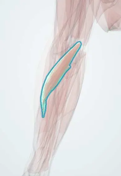
Pronator teres !
The pronator teres crosses the upper front of the forearm; it turns the palm down, and the median nerve passes between its two heads.
Opens in the Durgical app, no sign-up needed.
What it is
A short muscle from the inner elbow across to the middle of the radius.
What it does
Turns the palm down and helps bend the elbow.
Key facts
- Origin
- Humeral head: medial epicondyle; ulnar head: coronoid process
- Insertion
- Middle of the lateral surface of the radius
- Action
- Pronates the forearm; helps flex the elbow
- Nerve
- Median nerve (C6–C7)
- Blood supply
- Ulnar artery (anterior ulnar recurrent), radial artery
Relations
- Superficial
- Bicipital aponeurosis and antebrachial fascia near the elbow; the radial artery crosses over its insertion on the radius
- Deep
- Its ulnar head separates the median nerve (between the heads) from the ulnar artery, which passes deep to the ulnar head; flexor digitorum superficialis (FDS) and brachialis lie behind it
- Medial
- Flexor carpi radialis, the next muscle of the superficial flexor layer, sharing the common flexor tendon
- Lateral
- Brachioradialis, across the cubital fossa, which holds the biceps tendon, the brachial artery and the median nerve
How to find it
With the elbow bent to a right angle and the forearm pronated against resistance, it is felt as a firm oblique ridge running from just above the medial epicondyle down and outward toward the middle of the radius, forming the inner edge of the cubital fossa.
Examination
- Pronator compression test: firm pressure over the proximal muscle that brings on tingling in the median territory suggests pronator syndrome
- Resisted pronation while the elbow is slowly straightened: forearm ache with median paresthesias points to compression between its heads
On imaging
Axial MRI shows its humeral and ulnar heads in the proximal forearm, with the median nerve passing between them. Denervation oedema in the pronator teres and the forearm flexors suggests median nerve compression near the elbow; oedema in FPL, FDP to the index and pronator quadratus with a normal pronator teres points to the AIN. Ultrasound shows the nerve between the heads.
Clinical notes
- Pronator syndrome: aching in the front of the forearm and median paresthesias that include the palm (the palmar cutaneous branch is involved), unlike carpal tunnel syndrome; night symptoms are less typical
- Weakness of pronator teres places a median nerve lesion at or above the elbow; normal strength with a weak FPL points lower
- Classic tendon transfer for a permanent radial nerve palsy: pronator teres to the extensor carpi radialis brevis (ECRB) to restore wrist extension
Anatomical variants
- The ulnar head may be small or absent (present in roughly four of five limbs in one dissection series); the median nerve then runs deep to the whole muscle
- A supracondylar process of the humerus with the ligament of Struthers can give the muscle an extra origin and trap the median nerve and brachial artery
Development
Part of the ventral (flexor-pronator) mass of the forearm, which gathers into the common flexor origin on the medial epicondyle; the median nerve supplies this mass except flexor carpi ulnaris and the ulnar half of FDP.
Related conditions
Test yourself
Think of the answer, then tap the question to see it.
Where does the median nerve enter the forearm in relation to the pronator teres?
Between its humeral and ulnar heads, the first compression site of pronator syndrome.
How does pronator syndrome differ from carpal tunnel syndrome?
Forearm ache with median paresthesias that include the palm (palmar cutaneous branch), and night symptoms are less typical.
How is the proximal fragment displaced in a radial shaft fracture above the pronator teres insertion?
It is supinated by the biceps and supinator, while the distal fragment is pronated.
Which tendon transfer uses the pronator teres in a permanent radial nerve palsy?
Pronator teres to the extensor carpi radialis brevis (ECRB), to restore wrist extension.
Educational information, not medical advice. For any symptom, see a health professional.
Its names
- Official Latin (TA2)
- pronator teres TA2 2478
- Official Spanish (DeCS)
- Músculo pronador redondo
- English (TA2)
- Pronator teres
Related
Sources: Terminologia Anatomica (TA2); Anatomy, Shoulder and Upper Limb, Pronator Teres.
Spotted a mistake?We are always improving for the community. Tell us what is wrong and our team will review it.Report a mistake
Representative model, for educational purposes only.