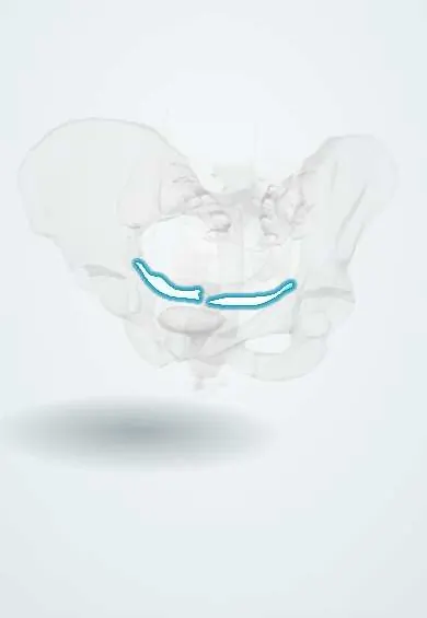
Reproductive system · Pelvis · Female
Cardinal ligament !
The cardinal ligament is dense connective tissue on each side of the cervix, around its vessels; it is the main side support of the cervix and upper vagina.
Opens in the Durgical app, no sign-up needed.
What it is
Thick connective tissue at the base of the broad ligament, from the cervix and upper vagina to the pelvic side wall.
What it does
The main side support of the cervix. The uterine artery runs in it and crosses over the ureter here.
Key facts
- Attaches
- Lateral cervix and upper vagina to the lateral pelvic wall (at the base of the broad ligament)
- Function
- Main lateral support of the cervix and vaginal vault (level I support); contains the uterine vessels, which cross over the ureter here
Relations
- Medial
- Cervix and the lateral fornix of the upper vagina
- Lateral
- Lateral pelvic wall: fascia over the obturator internus and the internal iliac vessels
- Above
- Base of the broad ligament
- Behind
- Uterosacral ligament, with which it blends at the cervix
- Below
- Levator ani of the pelvic floor
- Contents
- Uterine artery and uterine veins, vaginal artery, the ureters (in the ureteric tunnel), fibres of the inferior hypogastric plexus and lymphatics to obturator and iliac nodes
How to find it
Not felt as a separate band. At radical hysterectomy it is defined by opening the paravesical space in front and the pararectal space behind; the tissue left between them, running from the cervix to the side wall with the ureter passing through, is the cardinal ligament.
Examination
- POP-Q point C (cervix or vault) during straining measures failure of level I support: the further it comes down toward or past the hymen, the weaker the support
On imaging
On axial T2 MRI it appears as strands of tissue and vessels running from the cervix to the pelvic side wall. Its key role is cervical cancer staging: an intact dark ring of cervical stroma argues against parametrial invasion, while tumour reaching into this tissue upstages the disease, and hydronephrosis signals side wall spread. Dynamic MRI shows apical descent in prolapse.
Common problems
- Weakened supports allow prolapse.
- Endometriosis often grows on the uterosacral ligaments.
How it is diagnosed
Examination and ultrasound; seen directly in laparoscopy.
Treatment
- They are divided step by step in a hysterectomy.
Clinical notes
- At simple hysterectomy the uterine pedicle is clamped close to the cervix; clamping wide, out in the cardinal ligament, puts the ureter at risk
- The Manchester repair for prolapse shortens the cardinal ligaments and amputates part of the cervix
Memory aid
"Water under the bridge": the ureter (water) passes under the uterine artery (bridge) where both cross the cardinal ligament.
Related conditions
Test yourself
Think of the answer, then tap the question to see it.
What is the cardinal ligament made of?
Not a discrete cord but condensed connective tissue around the vessels and nerves of the cervix (paracervix), at the base of the broad ligament.
Which artery and which tubular structure cross within the cardinal ligament, and how?
The uterine artery crosses above the ureter about 2 cm lateral to the cervix; the ureter then runs through a tunnel in the ligament to the bladder.
What happens when the cardinal ligament is stretched or torn?
The cervix descends: uterine prolapse, or vault prolapse after hysterectomy (loss of level I support).
Why does wide clamping in the cardinal ligament during simple hysterectomy endanger the ureter?
The ureter lies about 2 cm lateral to the cervix there, so the uterine pedicle is clamped close to the cervix.
Educational information, not medical advice. For any symptom, see a health professional.
Its names
- Official Latin (TA2)
- ligamentum cardinale TA2 3839
- Official Spanish (DeCS)
- Ligamento cardinal
- English (TA2)
- Cardinal ligament
- Also called
- transverse cervical ligament · lateral cervical ligament · Mackenrodt's ligament
Related
Sources: Terminologia Anatomica (TA2); Anatomy, Abdomen and Pelvis: Cardinal Ligaments (Mackenrodts, Transverse Cervical, or Lateral Cervical Ligaments).
Spotted a mistake?We are always improving for the community. Tell us what is wrong and our team will review it.Report a mistake
Representative model, for educational purposes only.