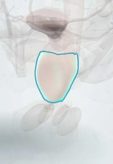
Pelvic organ prolapse !
Pelvic organ prolapse is the sagging of the bladder, uterus or rectum into or beyond the vagina because their muscle and tissue support has weakened.
Updated 2026-10-29
Structures involved
Tap one to see what it is and open it in 3D.
- Vagina
- Body of uterus
- Urinary bladder
- Rectum
- Uterosacral ligament
- Cardinal ligament
- Pubococcygeus
- Iliococcygeus
- Perineal body
In the clinical exam
Why it happens
What it is
Descent of one or more pelvic organs (bladder, uterus or vaginal vault, rectum or small bowel) into or beyond the vagina because their muscular and connective tissue support has weakened.
Who gets it
Very common in parous and older women, though many have no symptoms. It is one of the main reasons for gynaecological surgery in older women, and demand grows as the population ages.
Causes
- Damage to the levator ani muscle and its nerves during vaginal birth
- Weakening or tearing of the endopelvic fascia and the uterosacral and cardinal ligaments
- Long-term raised intra-abdominal pressure
- Loss of oestrogen and collagen with age and menopause
Risk factors
- Vaginal childbirth, especially several births, large babies or instrumental delivery
- Increasing age and menopause
- Obesity
- Chronic constipation and straining
- Chronic cough or heavy lifting
- Previous hysterectomy or prolapse surgery
- Connective tissue disorders and family history
Pathophysiology
The levator ani and connective tissue at three levels hold the pelvic organs: uterosacral and cardinal ligaments at the apex, lateral vaginal attachments, and the perineal body below. When the levator is damaged, the genital hiatus widens and the ligaments and fascia take the load until they stretch or tear, so the vaginal walls and the organs behind them descend.
Classification
- Anterior compartment: cystocele (bladder) or cystourethrocele
- Apical compartment: uterine prolapse or vaginal vault prolapse after hysterectomy
- Posterior compartment: rectocele (rectum) or enterocele (small bowel)
- Staging by the POP-Q system (stage 0–IV); older systems such as Baden–Walker are still used
- Procidentia: complete eversion of the uterus or vagina outside the introitus
Symptoms and signs
- Feeling of a bulge, lump or something coming down in the vagina
- Pelvic heaviness or dragging, worse at the end of the day or after standing
- Visible or palpable tissue at the vaginal opening
- Urinary symptoms: weak or interrupted stream, incomplete emptying, frequency, urgency or leakage
- Bowel symptoms: incomplete emptying, needing to press on the vagina or perineum to pass stool
- Sexual discomfort; many women have no symptoms
Diagnosis
- Clinical
- Pelvic examination at rest and while straining (supine and, if needed, standing) with a speculum blade to assess each compartment; POP-Q measurements; checks for stress incontinence (including with the prolapse reduced) and pelvic floor strength.
- Tests
- Urinalysis; post-void residual volume; urodynamic studies before surgery in selected women; kidney function if retention or procidentia is present.
- Imaging
- Usually not needed. Ultrasound measures residual urine or looks for hydronephrosis; pelvic floor ultrasound, defecography or MRI in complex or recurrent cases.
Differential diagnosis
- Cervical elongation
- Urethral diverticulum
- Gartner duct or other vaginal cyst
- Rectal prolapse
- Vaginal or cervical tumour
Goal of treatment
Relieve symptoms and restore bladder, bowel and sexual function; no treatment is needed for prolapse that causes no symptoms.
Treatment
- Follow-up
- Watchful waiting for women without symptoms or with mild symptoms who prefer no treatment.
- Rehabilitation
- Supervised pelvic floor muscle training, which can improve symptoms and stop mild prolapse from getting worse.
- Lifestyle
- Weight loss, treating constipation and chronic cough, and avoiding repeated heavy lifting.
- Conservative
- Vaginal pessary (ring, Gellhorn and other shapes), fitted and reviewed regularly; useful for women who prefer to avoid or are unfit for surgery.
- Medical
- Vaginal oestrogen in postmenopausal women to improve tissue quality and pessary tolerance.
- Surgical
- Native tissue repair (anterior or posterior colporrhaphy), apical suspension (sacrospinous or uterosacral fixation), sacrocolpopexy, hysterectomy in selected cases, or colpocleisis for women who no longer want vaginal intercourse.
Complications
- Urinary retention and recurrent urinary tract infection
- Hydronephrosis with severe prolapse
- Ulceration of exposed vaginal or cervical tissue
- Pessary problems: discharge, erosion, rarely fistula
- After surgery: recurrence, new stress incontinence, mesh complications, dyspareunia
Prognosis
Prolapse may stay stable for years and sometimes improves. Pessaries and surgery relieve symptoms in most women, but recurrence after surgery is common, particularly in the anterior compartment.
Prevention
- Pelvic floor muscle training, including after childbirth
- Healthy weight
- Preventing constipation and chronic straining
- Stopping smoking (chronic cough)
Test yourself
Think of the answer, then tap the question to see it.
Which muscle complex is the main active support of the pelvic organs?
The levator ani (pubococcygeus, puborectalis and iliococcygeus), whose injury in vaginal birth widens the genital hiatus.
Which organ descends in a cystocele and which in a rectocele?
The bladder in a cystocele (anterior wall); the rectum in a rectocele (posterior wall).
What is the most widely used objective staging system for pelvic organ prolapse?
The POP-Q (Pelvic Organ Prolapse Quantification) system, stages 0 to IV.
What is occult stress incontinence in prolapse?
Leakage that appears only when the prolapse is reduced, because the prolapse kinks the urethra; it can emerge after repair.
What is the main non-surgical device for symptomatic prolapse?
A vaginal pessary, combined in postmenopausal women with vaginal oestrogen and regular review.
Related conditions
- Stress urinary incontinence
- Rectal prolapse
- Obstetric perineal tears and anal sphincter injury
- Urinary tract infection (acute cystitis)
Educational information, not medical advice. For any symptom, see a health professional.
Sources: Pelvic Organ Prolapse (StatPearls (NCBI Bookshelf), 2025).
Spotted a mistake?We are always improving for the community. Tell us what is wrong and our team will review it.Report a mistake
Representative model, for educational purposes only.