Cardiovascular system · Upper limb
Circumflex humeral arteries !
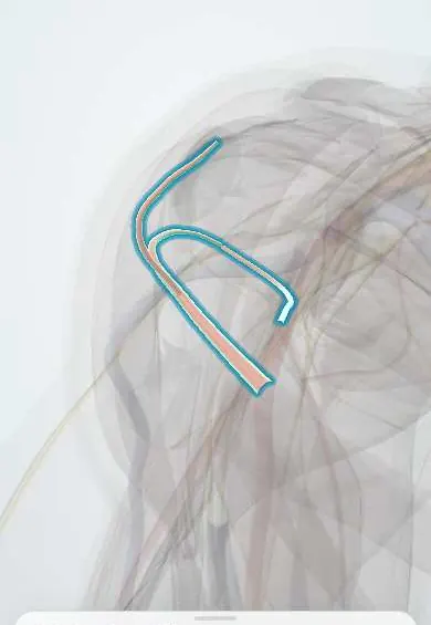
The circumflex humeral arteries wrap around the neck of the humerus; they feed the humeral head and deltoid, and a fracture here can cut that supply.
Official name: Anterior and posterior circumflex humeral arteries
Opens in the Durgical app, no sign-up needed.
What it is
Two arteries that circle the neck of the humerus.
What it does
Feed the humeral head and the deltoid; a fracture here can cut off the head's blood supply.
Key facts
- Origin
- 3rd part of the axillary artery
- Branches
- Anterior (to the humeral head via the arcuate artery); posterior (with the axillary nerve through the quadrangular space)
- Supplies
- Humeral head, deltoid, shoulder joint
Relations
- Course
- Both arise from the third part of the axillary artery near the lower border of subscapularis; the anterior runs laterally in front of the surgical neck, deep to coracobrachialis and the biceps short head; the posterior runs backward through the quadrangular space and winds round the neck
- Deep
- Surgical neck of the humerus and the shoulder capsule
- Superficial
- The deltoid, which the posterior artery supplies from behind together with the axillary nerve
How to find it
Not palpable. The posterior artery is found with the axillary nerve in the quadrangular space, approached from behind below the rear deltoid; the anterior artery and its two veins are met at the lower border of subscapularis in the deltopectoral approach.
On imaging
Not seen on radiographs, but a displaced fracture of the proximal humerus, above all through the anatomical neck, warns that the supply of the humeral head may be cut, with later avascular necrosis seen on MRI. CT angiography looks for arterial injury after fracture-dislocation. On MRI or ultrasound the posterior artery marks the quadrangular space and the axillary nerve.
Clinical notes
- Deltopectoral approach: the anterior circumflex humeral artery and its two veins (the "three sisters") mark the lower border of subscapularis and are tied or coagulated when that tendon is released
- Quadrangular space syndrome: fibrous bands or enlarged muscles compress the axillary nerve and posterior circumflex humeral artery in overhead athletes; the artery can also form an aneurysm or clot that sends emboli to the hand
- Proximal humerus fractures through the anatomical neck, or with little of the medial calcar left on the head, carry a higher risk of avascular necrosis of the head
Anatomical variants
- The posterior artery may arise from the subscapular artery, from a common trunk with the anterior one or from the deep brachial artery
Related conditions
Test yourself
Think of the answer, then tap the question to see it.
Which circumflex humeral artery passes through the quadrangular space, and with which nerve?
The posterior circumflex humeral artery, with the axillary nerve.
Why can a displaced surgical neck fracture threaten the humeral head?
Both circumflex humeral arteries wrap around the surgical neck and supply the head; a fracture there can cut off that supply.
What are the «three sisters» in the deltopectoral approach?
The anterior circumflex humeral artery and its two veins, marking the lower border of subscapularis; they are tied or coagulated.
Educational information, not medical advice. For any symptom, see a health professional.
Its names
- Official Latin (TA2)
- arteria circumflexa anterior humeri TA2 4630
- Official Spanish (DeCS)
- Arterias circunflejas anterior y posterior del húmero
- English (TA2)
- Anterior and posterior circumflex humeral arteries
Related
Sources: Terminologia Anatomica (TA2); Anatomy, Shoulder and Upper Limb, Anterior Humeral Circumflex Artery; Anatomy, Head and Neck, Posterior Humeral Circumflex Artery.
Spotted a mistake?We are always improving for the community. Tell us what is wrong and our team will review it.Report a mistake
Representative model, for educational purposes only.