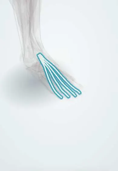
Toe extensor tendons !
The toe extensor tendons are four tendons crossing the top of the foot to the four smaller toes; they lift the toes.
Official name: Tendons of extensor digitorum longus
Opens in the Durgical app, no sign-up needed.
What it is
Four slim tendons across the top of the foot, from the extensor digitorum longus to the four smaller toes.
What it does
Lift the four smaller toes and help lift the foot.
Key facts
- Attaches
- Extensor digitorum longus: four tendons under extensor retinacula to dorsal expansions of lateral four toes (middle and distal phalanges)
- Function
- Extend lateral four toes; assist ankle dorsiflexion
- Tight in
- Toe flexion with ankle plantarflexion
Relations
- Course
- Pass under the superior extensor retinaculum, then through the lateral loop of the inferior extensor retinaculum in a shared sheath with the fibularis tertius, and fan out across the dorsum to the four lesser toes
- Medial
- Extensor hallucis longus tendon, with the dorsalis pedis artery and deep fibular nerve between them
- Lateral
- Fibularis tertius tendon, heading to the fifth metatarsal
- Deep
- Extensor digitorum brevis belly, whose tendons join those to the second to fourth toes, and the tarsal bones
- Superficial
- Dorsal venous arch and branches of the superficial fibular nerve
How to find it
Extending the toes against resistance raises four cords fanning across the top of the foot, lateral to the extensor hallucis longus tendon; at the ankle they lie together just lateral to it.
Examination
- Resisted extension of the lesser toes: weakness, together with foot drop, points to the deep fibular nerve or the L5 root
On imaging
Ultrasound is the first choice: the tendons run in front of the ankle under the extensor retinacula and fan out over the dorsum. Dynamic scanning shows a tear or the gap after a laceration, and tenosynovitis as fluid around the tendon in its sheath. MRI is kept for unclear cases. Radiographs only help to look for a fracture or a foreign body after a wound on the dorsum.
Common problems
- Cuts on the top of the foot can injure them; tight shoes can irritate them.
Clinical notes
- Cuts on the dorsum can divide these tendons and the nearby branches of the superficial fibular nerve together
- Anterior tarsal tunnel syndrome: the deep fibular nerve is compressed beneath the inferior extensor retinaculum, close to these tendons
- Claw and hammer toes reflect an imbalance in which these long extensors hold the MTP joints extended while the long and short flexors bend the interphalangeal joints
Anatomical variants
- Fibularis tertius, often regarded as a fifth slip of the extensor digitorum longus, can be small or absent; extra tendon slips are common
Memory aid
Front of the ankle, medial to lateral: "The Hospitals Are Not Dirty Places" (Tibialis anterior, Hallucis longus, Artery, Nerve, Digitorum longus, Peroneus tertius).
Test yourself
Think of the answer, then tap the question to see it.
What is the order of structures across the front of the ankle, medial to lateral?
Tibialis anterior, extensor hallucis longus, anterior tibial artery and deep fibular nerve, toe extensor tendons, fibularis tertius.
Where do the toe extensor tendons insert, and what do they do?
On the dorsal expansions of the lateral four toes (middle and distal phalanges); they extend these toes and assist ankle dorsiflexion.
What early sign of anterior compartment syndrome involves these tendons' muscle?
Pain on passive toe flexion, which stretches the extensor digitorum longus.
Educational information, not medical advice. For any symptom, see a health professional.
Its names
- Official Latin (TA2)
- extensor longus digitorum TA2 2645
- Official Spanish (DeCS)
- Tendones del músculo extensor largo de los dedos DeCS D013710
- English (TA2)
- Tendons of extensor digitorum longus
- Also called
- extensor digitorum longus muscle
Related
- Toe extensor tendon
- Extensor digitorum brevis
- Second metatarsal
- Third metatarsal
- Fourth metatarsal
- Second toe
- Third toe
- Fourth toe
Sources: Terminologia Anatomica (TA2); DeCS; Anatomy, Bony Pelvis and Lower Limb: Leg Anterior Compartment.
Spotted a mistake?We are always improving for the community. Tell us what is wrong and our team will review it.Report a mistake
Representative model, for educational purposes only.