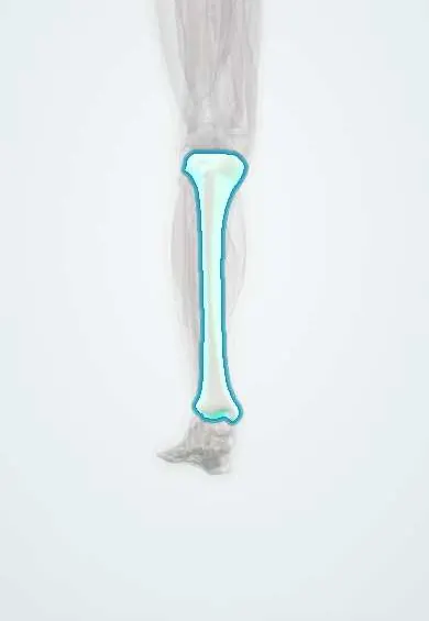
Medial tibial stress syndrome (shin splints) !
Medial tibial stress syndrome, or shin splints, is exercise-related pain along the inner edge of the shin bone from repeated loading.
Updated 2026-10-29
Structures involved
Tap one to see what it is and open it in 3D.
- Tibia
- Soleus
- Tibialis posterior
- Crural fascia
- Flexor digitorum longus
- Gastrocnemius (inner head)
- Interosseous membrane
- Navicular
Why it happens
What it is
Exercise-related pain along the inner (posteromedial) border of the lower two-thirds of the tibia, known as shin splints. It is a bone stress reaction caused by repeated loading.
Who gets it
One of the most common overuse injuries in runners, military recruits and dancers, and in sports with jumping.
Causes
- Repeated impact loading of the tibia
- Sudden increase in running distance, speed or frequency
- Training on hard surfaces or with worn footwear
Risk factors
- Female sex
- Higher body mass index
- Excessive foot pronation or navicular drop
- Previous medial tibial stress syndrome
- Little running experience
- Reduced hip range of motion in some studies
Pathophysiology
The exact mechanism is debated. One view is traction on the periosteum by the soleus, flexor digitorum longus, tibialis posterior and crural fascia; the more accepted view is bone overload, with remodeling that cannot keep up with repeated bending. It sits on a continuum with tibial stress fracture.
Symptoms and signs
- Diffuse aching pain along the inner border of the shin, over several centimeters
- Pain at the start of exercise that may ease while running, then returns after
- In later stages, pain with less activity and at rest
- Diffuse tenderness along the posteromedial tibia
Diagnosis
- Clinical
- Diagnosis is clinical: exercise-induced pain and diffuse tenderness over the posteromedial tibia, without features of other causes.
- Tests
- Compartment pressure testing if chronic exertional compartment syndrome is suspected; bone health tests in recurrent bone stress injury.
- Imaging
- X-rays are usually normal. MRI separates periosteal and marrow edema from a fracture line; bone scan is an alternative.
Differential diagnosis
- Tibial stress fracture
- Chronic exertional compartment syndrome
- Popliteal artery entrapment syndrome
- Muscle strain or tendinopathy
- Nerve entrapment
- Bone tumor or infection
Goal of treatment
Let the bone adapt by reducing load, keep fitness with low-impact activity and return gradually to running while correcting risk factors.
Treatment
- Conservative
- Relative rest from impact, ice, and cross-training such as cycling or swimming.
- Medical
- Short courses of simple analgesics or NSAIDs for pain.
- Rehabilitation
- Graded return-to-run plan; calf, foot and hip strengthening; review of running technique.
- Conservative
- Suitable footwear and, for excessive pronation, orthoses; evidence for these and for shockwave therapy is limited.
- Surgical
- Rarely, posteromedial fasciotomy for long-standing pain that fails rehabilitation.
Complications
- Progression to tibial stress fracture
- Recurrence with early return to training
- Long time away from sport
Prognosis
Most athletes recover with load management, though recovery can take weeks to months and recurrence is common.
Prevention
- Gradual increases in training load
- Rest days and varied surfaces
- Strength training of the calf and hip
Test yourself
Think of the answer, then tap the question to see it.
Where is the pain located in medial tibial stress syndrome?
Along the posteromedial border of the lower two-thirds of the tibia, over a diffuse area.
Which finding suggests a tibial stress fracture rather than shin splints?
Focal point tenderness with night or rest pain, rather than diffuse tenderness along the shin.
How does chronic exertional compartment syndrome differ from shin splints?
It causes tight, burning pain and sometimes numbness that builds during exercise and settles with rest; compartment pressure testing confirms it.
Which imaging test separates stress reaction from stress fracture?
MRI, which shows periosteal or marrow edema with or without a fracture line.
What are two risk factors for medial tibial stress syndrome?
Female sex, higher body mass index, excessive pronation, previous episodes or a sudden rise in training load.
Related conditions
- Acute compartment syndrome
- Tibial shaft fracture
- Metatarsal fractures (including stress fractures)
- Flatfoot (pes planus)
- Achilles Tendinopathy
Educational information, not medical advice. For any symptom, see a health professional.
Sources: Medial Tibial Stress Syndrome (StatPearls (NCBI Bookshelf), 2025).
Spotted a mistake?We are always improving for the community. Tell us what is wrong and our team will review it.Report a mistake
Representative model, for educational purposes only.