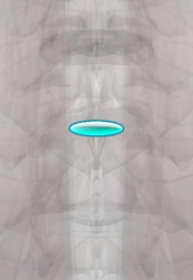
L3–L4 nucleus !
The nucleus pulposus is the soft, gel-like centre of the L3-L4 disc in the lower back; it works like a water cushion that spreads the load.
Official name: L3-L4
Opens in the Durgical app, no sign-up needed.
What it is
The soft, gel-like centre (nucleus pulposus) of the L3–L4 disc.
What it does
Works like a water cushion: it turns pressure from above into outward push on the fibre ring, spreading the load. It is under the most pressure when you bend forward and lift.
Key facts
- Where
- Centre of the L3–L4 disc, slightly toward the back, inside the annulus fibrosus
- Function
- Notochord-derived gel (about 70–90% water, proteoglycans); turns axial load into annulus tension; herniates posterolaterally toward the L4 root
Relations
- Behind
- Annulus and posterior longitudinal ligament, then the dural sac; the L4 root runs in the lateral recess
- Lateral
- The L3 root and its ganglion in and just outside the foramen, where a far-lateral fragment lands
How to find it
The line joining the tops of the iliac crests (Tuffier line) crosses about the L4 spinous process or the L4–L5 space, so the L3–L4 gap is the one just above it, a common space for lumbar puncture.
Examination
- L4 root: weak ankle dorsiflexion (tibialis anterior), numb inner shin, reduced knee jerk (L3–L4)
On imaging
Axial MRI cuts must reach beyond the pedicle: a far-lateral fragment sits outside the foramen next to the dorsal root ganglion and is easy to miss on midline sagittal images.
Common problems
- Squeezes out through a tear in the ring (herniation), usually backward and to one side, toward a nerve root.
- Loses water with age, so the disc gets thinner and stiffer.
How it is diagnosed
On MRI a healthy nucleus is bright on T2 images; a worn one turns dark ('black disc').
Treatment
- In a microdiscectomy only the loose piece that has pushed out is removed; most of the disc stays.
Clinical notes
- A far-lateral fragment is reached through a paramedian muscle-splitting approach between the multifidus and the longissimus (Wiltse), rather than through the midline interlaminar route
- Adjacent segment disease: after fusion of L4–L5 or L4–S1, movement and load shift to the next free disc, so L3–L4 often degenerates or herniates later
- In lateral transpsoas interbody fusion the lumbar plexus lies further forward within the psoas at the lower lumbar levels, so the femoral nerve is more at risk at L3–L4 and L4–L5 than higher up
Related conditions
Test yourself
Think of the answer, then tap the question to see it.
Toward which root does a posterolateral herniation of the L3–L4 nucleus usually go?
The L4 root; a far-lateral fragment reaches the L3 root instead.
What picture does a far-lateral L3–L4 nucleus fragment give?
Front-of-thigh pain and a weak knee jerk from the L3 root, instead of the expected L4 picture.
Why must axial MRI cuts reach beyond the pedicle to find a far-lateral L3–L4 nucleus fragment?
The fragment sits outside the foramen next to the dorsal root ganglion and is easy to miss on midline sagittal images.
Which approach reaches a far-lateral L3–L4 nucleus fragment?
A paramedian muscle-splitting approach between the multifidus and the longissimus (Wiltse), rather than the midline interlaminar route.
Educational information, not medical advice. For any symptom, see a health professional.
Its names
- Official Latin (TA2)
- nucleus pulposus L3-L4 TA2 1686
- Official Spanish (DeCS)
- Núcleo pulposo L3-L4 DeCS D000070614
- English (TA2)
- L3-L4
Related
Sources: Terminologia Anatomica (TA2); DeCS; Anatomy, Back, Nucleus Pulposus; Anatomy, Back, Intervertebral Discs; 7.3 The Vertebral Column (OpenStax (Rice University), 2022) · adapted, CC BY 4.0.
Spotted a mistake?We are always improving for the community. Tell us what is wrong and our team will review it.Report a mistake
Representative model, for educational purposes only.