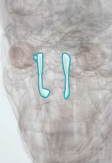
Tear drainage !
The tear drainage system is a set of tiny channels, a sac and a duct that carry tears from the inner corner of the eye into the nose.
Official name: Lacrimal canaliculus
Opens in the Durgical app, no sign-up needed.
What it is
Tiny channels from the inner corner of the eyelids into a sac and a duct that opens into the nose.
What it does
Drain tears into the nose; a blocked duct causes a watery eye.
Key facts
- Where
- Puncta -> canaliculi -> lacrimal sac -> nasolacrimal duct -> inferior meatus
- Function
- Drains tears into the nose
Relations
- Course
- From the puncta on the medial lid margins, the canaliculi run medially behind the medial canthal tendon to the sac; the nasolacrimal duct then descends in a bony canal of the maxilla, slightly backward and outward, to the inferior meatus
- Medial
- Between the sac and the nasal cavity, at the front of the middle meatus, lie the lacrimal bone and the maxilla's frontal process, and sometimes anterior ethmoid cells
- In front
- Medial palpebral ligament across the upper part of the sac; skin, with the angular vein a little medially
- Behind
- Lacrimal part of orbicularis oculi (Horner muscle), attached to the posterior lacrimal crest; orbital septum
- Lateral
- Along the nasolacrimal duct, the maxillary sinus beside its bony canal
How to find it
The puncta sit on small raised papillae at the inner end of each lid margin, turned toward the eye, and show when the lid is drawn gently away. The sac lies in the bony groove at the inner corner of the eye, behind the medial canthal tendon, which stands out as a cord when the lids are pulled outward. The lower opening of the duct is seen under the inferior concha on nasal endoscopy.
Examination
- Fluorescein dye disappearance test: dye placed in both lower fornices clears within a few minutes in a normal system; dye retained on one side suggests a block (useful in children)
- Lacrimal irrigation through a punctum: fluid reaching the nose means the system is open; reflux from the same punctum points to a canalicular block, reflux from the opposite punctum to a block in the common canaliculus, sac or duct
- Pressure over the sac that brings mucus or pus out of the puncta shows a blocked nasolacrimal duct with a chronically infected sac
On imaging
Dacryocystography (contrast injected into a canaliculus) outlines the level of a block, and dacryoscintigraphy shows how well tears drain. CT is used after naso-orbito-ethmoid fractures or when a tumour of the sac is suspected.
Common problems
- Blocked tear duct: a constantly watery eye; common in babies and older adults.
- Dacryocystitis: an infected, swollen tear sac by the nose.
How it is diagnosed
Flushing the duct with saline and dye tests.
Treatment
- Massage in babies (most open by themselves).
- Probing, or surgery to make a new path into the nose (dacryocystorhinostomy).
Clinical notes
- Congenital nasolacrimal duct obstruction: watering and sticky discharge from the first weeks of life; most open on their own within the first year, and persistent cases are probed
- Dacryocystitis: a red, tender swelling below the medial canthal tendon; a mass above the tendon is atypical and raises concern for a tumour of the sac
- Dacryocystorhinostomy (DCR) bypasses a blocked duct by joining the sac to the nasal cavity through an opening in the lacrimal bone and frontal process of the maxilla, by an external or endoscopic route
- A watery eye in an adult may come from too many tears (irritation, ectropion) or poor drainage; a punctum turned away from the eye is a common, simple cause in older people
Development
The duct forms from a solid cord of surface ectoderm buried along the nasolacrimal groove, between the maxillary and lateral nasal prominences; it hollows out from above downward and the lower end opens last, which explains congenital obstruction.
Related conditions
Test yourself
Think of the answer, then tap the question to see it.
What is the path of tear drainage from the eye to the nose?
Puncta, canaliculi, lacrimal sac, nasolacrimal duct, then the inferior meatus under the inferior nasal concha.
Where is the usual site of congenital nasolacrimal duct obstruction, and how does it usually evolve?
The membrane at the lower end of the duct (valve of Hasner); most open on their own within the first year.
Where does dacryocystitis swell, and what does a mass above the medial canthal tendon suggest?
Below the medial canthal tendon, red and tender; a mass above the tendon is atypical and raises concern for a tumour of the sac.
What does a dacryocystorhinostomy (DCR) do?
It bypasses a blocked nasolacrimal duct by joining the lacrimal sac to the nasal cavity through an opening in the lacrimal bone and maxilla.
Educational information, not medical advice. For any symptom, see a health professional.
Its names
- Official Latin (TA2)
- canaliculus lacrimalis TA2 6855
- Official Spanish (DeCS)
- Vías lagrimales DeCS D009301
- English (TA2)
- Lacrimal canaliculus
- Also called
- tear duct · ductus lacrimales · ductuli lacrimales
Related
Sources: Terminologia Anatomica (TA2); DeCS; Anatomy, Head and Neck: Eye Nasolacrimal; Anatomy, Head and Neck: Eye Lacrimal Duct; Anatomy of the Human Body (20th ed.): The Accessory Organs of the Eye (Lea & Febiger (Gray's Anatomy, 1918), 1918) · adapted, Public domain.
Spotted a mistake?We are always improving for the community. Tell us what is wrong and our team will review it.Report a mistake
Representative model, for educational purposes only.