Conditions · Ophthalmology and ENT
Dacryocystitis and nasolacrimal duct obstruction !
Blockage of the tear drainage duct makes the eye water constantly; if the tear sac behind it gets infected, a painful red lump forms beside the nose.
Updated 2026-11-01
Structures involved
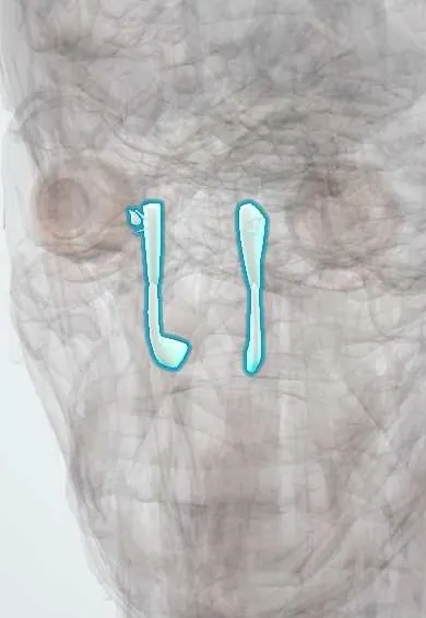
Tap one to see what it is and open it in 3D.
- Tear drainage
- Eyelids
- Lacrimal gland
- Lacrimal bones
- Nasal cavity
- Inferior nasal conchae
- Maxillae
- Cavernous sinus
Why it happens
- Patterns of acute inflammation: serous, fibrinous, purulent, abscess, ulcer
- Conjunctiva, eyelid glands and lacrimal gland
What it is
Nasolacrimal duct obstruction is a partial or complete blockage of the passage that carries tears from the lacrimal sac into the nose, causing tearing (epiphora). Dacryocystitis is infection and inflammation of the lacrimal sac that develops because stagnant tears behind the blockage become colonised. It may be acute, with a tender swelling at the inner corner of the eye, or chronic, with tearing and mucopurulent discharge.
Who gets it
Congenital obstruction is common in newborns, affecting a few percent of babies, and most open spontaneously during the first year. Acquired obstruction and dacryocystitis are typical of adults over 40, more often women, whose duct is narrower. It is a frequent cause of chronic tearing in ophthalmology and primary care in Colombia.
Causes
- Congenital: a persistent membrane at the lower end of the duct (valve of Hasner)
- Acquired primary: idiopathic inflammation and fibrosis of the duct with age
- Acquired secondary: nasal and facial fractures, sinus or nasal surgery, chronic sinusitis, granulomatous diseases (granulomatosis with polyangiitis, sarcoidosis), stones (dacryoliths) and tumours
- Infection of the sac: Staphylococcus aureus, streptococci, Haemophilus influenzae in children; staphylococci, pneumococcus and Pseudomonas in adults
Risk factors
- Female sex and age over 40
- Newborn period (congenital obstruction)
- Midface trauma, including nasal fracture
- Chronic rhinosinusitis and previous nasal or sinus surgery
- Systemic granulomatous disease
- Some topical and systemic medicines that scar the drainage system
Pathophysiology
Tears drain through the puncta and canaliculi into the lacrimal sac and then down the nasolacrimal duct to the inferior meatus beneath the inferior concha. When the duct narrows or closes, tears pool in the sac, overflow onto the cheek and become a culture medium. Bacterial infection inflames the sac wall; in acute disease this causes cellulitis around the sac, and pus can break through the skin (fistula) or spread into the orbit.
Classification
- Congenital nasolacrimal duct obstruction
- Acquired nasolacrimal duct obstruction: primary or secondary
- Acute dacryocystitis
- Chronic dacryocystitis (often with a mucocele of the sac)
- Congenital dacryocystocele in newborns (bluish swelling below the inner canthus)
Symptoms and signs
- Persistent tearing (epiphora) and tears running onto the cheek
- Mucous or purulent discharge, crusting of the lashes, often in one eye
- Pressure on the sac area pushes mucus or pus back out of the puncta (regurgitation test)
- Acute dacryocystitis: painful, red, tense swelling below the medial canthal tendon, beside the nose
- Fever and malaise in severe acute cases
- Infants: watery, sticky eye from the first weeks of life with a white, non-red eye
Diagnosis
- Clinical
- History and examination: medial canthal swelling, regurgitation of pus on pressing the sac, and the dye disappearance test (fluorescein remaining in the tear film suggests poor drainage). Irrigation of the drainage system by the ophthalmologist confirms obstruction once the acute infection has settled.
- Tests
- Gram stain and culture of discharge expressed from the puncta in acute cases. Blood tests and autoimmune serology only when systemic disease or toxicity is suspected.
- Imaging
- CT of the orbits and sinuses if orbital spread, trauma or a tumour is suspected. Nasal endoscopy and dacryocystography assess the anatomy before surgery.
Differential diagnosis
- Preseptal or orbital cellulitis
- Acute ethmoid sinusitis
- Hordeolum or chalazion
- Conjunctivitis (discharge without sac swelling or tearing from obstruction)
- Congenital glaucoma in an infant with tearing and light sensitivity (enlarged, cloudy cornea)
- Lacrimal sac tumour
Goal of treatment
Control infection, prevent orbital spread, and restore tear drainage so that infection does not recur.
Treatment
- Medical
- Acute dacryocystitis: systemic antibiotics against gram-positive organisms, warm compresses and pain relief; intravenous antibiotics and admission when there is orbital cellulitis, systemic illness or failure of oral treatment.
- Procedure
- Drainage of a pointing lacrimal sac abscess when needed; the duct is not probed during acute infection.
- Conservative
- Congenital obstruction: observation with lacrimal sac massage (downward pressure over the sac) and lid hygiene, since most resolve in the first year.
- Procedure
- Infants whose obstruction persists: probing of the nasolacrimal duct, with or without silicone intubation.
- Surgical
- Adults with established obstruction or chronic dacryocystitis: dacryocystorhinostomy, which creates a new opening from the lacrimal sac into the nose (external or endoscopic).
Complications
- Orbital cellulitis and orbital abscess
- Lacrimal fistula to the skin
- Chronic mucocele of the sac
- Meningitis or cavernous sinus thrombosis (rare)
- Infection after intraocular surgery if a chronically infected sac is not treated first
Prognosis
Congenital obstruction resolves on its own in most infants, and probing succeeds in most of the rest. Acute dacryocystitis responds to antibiotics, but recurrence is common unless the obstruction is relieved; dacryocystorhinostomy has a high success rate.
Prevention
- Prompt treatment of chronic sinusitis and nasal disease
- Assessment of tear drainage after midface fractures
- Treating a chronically infected sac before cataract or other eye surgery
Test yourself
Think of the answer, then tap the question to see it.
A woman of 60 has a painful red swelling below the medial canthus and pus on pressing it. Diagnosis?
Acute dacryocystitis.
Should the nasolacrimal duct be probed during acute dacryocystitis?
No; treat the infection first and relieve the obstruction later.
First-line management of congenital nasolacrimal duct obstruction?
Observation with lacrimal sac massage; most resolve by 12 months.
Definitive operation for adult nasolacrimal duct obstruction with recurrent dacryocystitis?
Dacryocystorhinostomy.
Which complication must be excluded if a patient with dacryocystitis develops proptosis and painful eye movements?
Orbital cellulitis.
Related conditions
- Orbital cellulitis
- Conjunctivitis
- Chalazion and hordeolum (stye)
- Chronic rhinosinusitis and nasal polyps
- Acute rhinosinusitis
- Granulomatosis with polyangiitis
- Sarcoidosis
- Nasal fracture and septal haematoma
- Bacterial meningitis
Educational information, not medical advice. For any symptom, see a health professional.
Sources: Dacryocystitis (EyeWiki, American Academy of Ophthalmology, 2026); Blocked tear duct (MedlinePlus, US National Library of Medicine, 2026).
Spotted a mistake?We are always improving for the community. Tell us what is wrong and our team will review it.Report a mistake
Representative model, for educational purposes only.