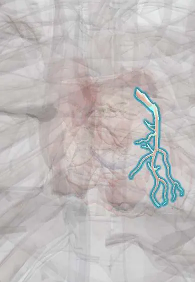
LAD (anterior descending artery) !
The left anterior descending artery runs down the front of the heart; it feeds the front of the left ventricle and most of the septum.
Official name: Anterior interventricular artery
Opens in the Durgical app, no sign-up needed.
What it is
The left anterior descending artery, running down the front groove between the ventricles, with diagonal and septal branches.
What it does
Feeds the front of the left ventricle and most of the septum. A sudden blockage near its start is sometimes called the 'widow-maker'.
Key facts
- Origin
- Left main coronary artery; runs in anterior interventricular groove to apex
- Branches
- Diagonal branches; septal perforators; conus branch
- Supplies
- Anterior LV wall, apex, anterior 2/3 of septum and bundle branches; strip of anterior RV
Relations
- Course
- From the left main coronary artery it passes to the left of the pulmonary trunk and descends in the anterior interventricular groove to the apex
- Superficial
- Epicardial fat and visceral pericardium; the great cardiac vein ascends alongside it in the groove
- Deep
- Interventricular septum, which its septal perforators enter at right angles
How to find it
Seen on the front of the heart once the pericardium is opened: it runs in the anterior interventricular groove toward the apex, often buried in fat or under a muscle bridge, with the great cardiac vein beside it. Surgeons find it there to sew the internal thoracic artery graft, usually in its middle third.
On imaging
On coronary angiography the right and left anterior oblique cranial views separate it from its diagonal and septal branches; it is described as proximal (up to the first septal perforator or first diagonal), mid and distal segments. CT coronary angiography shows plaque and any myocardial bridge.
Common problems
- Coronary artery disease: fatty plaque narrows them, causing chest pain on exertion (angina).
- Heart attack: a plaque breaks open and a clot suddenly blocks the artery.
How it is diagnosed
ECG and troponin for a heart attack; stress test, CT coronary angiography or a catheter angiogram to see narrowings.
Treatment
- Medicines (aspirin, statins, blood-pressure control) and stopping smoking.
- Angioplasty and stent through a catheter.
- Bypass surgery (CABG) for several or complex blockages.
Clinical notes
- Revascularization is by stenting through a catheter or by a bypass graft, usually the left internal thoracic artery sewn to the mid LAD
- Large anterior infarcts can lead to an apical aneurysm with mural thrombus, rupture of the interventricular septum and pump failure
- In a chronic LAD occlusion, septal collaterals from the posterior descending artery often refill it
Anatomical variants
- Myocardial bridging: a segment, most often the mid LAD, runs within the heart muscle and is squeezed in systole; usually harmless, occasionally linked to ischemia
- A long 'wrap-around' LAD curls round the apex into the posterior groove, so its occlusion can also affect the inferior wall
Related conditions
- Familial hypercholesterolemia
- Familial dysbetalipoproteinemia (type III hyperlipoproteinemia)
- Familial combined hyperlipidaemia
- Cardiogenic shock
- Vasospastic (Prinzmetal) angina
- Myocardial Infarction (Heart Attack)
- Stable angina
Test yourself
Think of the answer, then tap the question to see it.
Where does the LAD run, and which branches does it give?
From the left main coronary artery down the anterior interventricular groove to the apex; it gives diagonal branches and septal perforators.
What ECG changes mark an anterior myocardial infarction from LAD occlusion?
ST elevation in V1–V4; a proximal («widow-maker») occlusion also involves the diagonals, with changes extending to I, aVL and V5–V6.
Which bypass graft to the LAD is the most durable?
The left internal thoracic (mammary) artery graft, the standard in coronary artery bypass surgery.
What is the Wellens pattern?
Deep symmetrical or biphasic T waves in V2–V3 after chest pain, pointing to a critical proximal LAD stenosis.
What is myocardial bridging, and which artery is most often involved?
A segment that runs within the heart muscle and is squeezed in systole, most often the mid LAD; usually harmless.
Educational information, not medical advice. For any symptom, see a health professional.
Its names
- Official Latin (TA2)
- arteria interventricularis anterior; ramus diagonalis arteriae interventricularis anterioris TA2 4143
- Official Spanish (DeCS)
- Arteria interventricular anterior DeCS D003331
- English (TA2)
- Anterior interventricular artery
- Also called
- anterior interventricular branch of left coronary artery · lateral branch of anterior interventricular artery · LAD · left anterior descending artery · ramus interventricularis arteriae coronariae sinistrae · ramus descendens anterior
Related
- Great cardiac vein
- Left ventricle
- Interventricular septum
- Bundle branches
- Left ventricle papillary muscles
Sources: Terminologia Anatomica (TA2); DeCS; Anatomy, Thorax, Heart Left Anterior Descending (LAD) Artery; 19.1 Heart Anatomy (OpenStax (Rice University), 2022) · adapted, CC BY 4.0.
Spotted a mistake?We are always improving for the community. Tell us what is wrong and our team will review it.Report a mistake
Representative model, for educational purposes only.