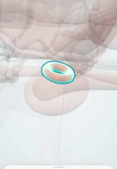
External urethral sphincter !
The external urethral sphincter is the ring of voluntary muscle around the urethra; it holds urine in when coughing, laughing or lifting.
Official name: Female external urethral sphincter
Opens in the Durgical app, no sign-up needed.
What it is
A ring of voluntary (striated) muscle around the membranous urethra, just below the apex of the prostate (rhabdosphincter).
What it does
Holds urine in by choice; after the prostate is removed it is the main muscle that keeps a man continent.
Key facts
- Origin
- Encircles the middle third of the urethra; compressor urethrae from the ischiopubic rami
- Insertion
- Ring around the urethra; compressor urethrae and urethrovaginal sphincter arch over the urethra and around the vagina
- Action
- Voluntary closure of the urethra (continence under effort)
- Nerve
- Perineal branch of pudendal nerve (S2–S4)
- Blood supply
- Internal pudendal artery branches
Relations
- Above
- The bladder neck with the internal (smooth) sphincter; in the male, the apex of the prostate
- Below
- The perineal membrane, with the superficial perineal pouch beneath
- Behind
- Female: the anterior vaginal wall. Male: the rectum, with the bulbourethral glands beside the membranous urethra
- Lateral
- The ischiopubic rami, from which the compressor urethrae arises, and the medial edges of levator ani
- Contents
- The middle third of the female urethra; in the male, the membranous urethra
How to find it
Not palpable. In women it surrounds the middle third of the urethra, behind the pubic symphysis and in front of the anterior vaginal wall. In men it is found in surgery at the membranous urethra just below the apex of the prostate, where it is preserved during radical prostatectomy.
Examination
- Cough stress test: with a comfortably full bladder, urine escaping from the meatus at the very moment of a cough shows stress incontinence; leakage that starts later suggests detrusor overactivity
- Urodynamics: a low abdominal leak point pressure or a low urethral closure pressure points to intrinsic sphincter deficiency rather than urethral hypermobility
On imaging
Transperineal or endovaginal ultrasound and MRI show it as a ring of striated muscle around the middle third of the urethra; thinning or a defect supports sphincter weakness.
Common problems
- Damage or weakness after a radical prostatectomy causes stress urinary incontinence, which usually improves over months.
How it is diagnosed
History, pad test and bladder diary; urodynamic tests sometimes.
Treatment
- Pelvic floor training before and after surgery; an artificial urinary sphincter or a male sling if leakage persists.
Clinical notes
- Pelvic floor muscle training aims to strengthen it together with levator ani; it is the usual first approach to stress incontinence
- Stretching of the pudendal nerve in a long or assisted vaginal birth can weaken it
- Intrinsic sphincter deficiency means the closing mechanism itself fails, unlike incontinence from urethral hypermobility
- Male: damaged in radical prostatectomy and in pelvic fractures that tear the membranous urethra
Development
In the fetus it forms a continuous sleeve of striated muscle from the bladder neck down to the perineal membrane. In the male the growing prostate thins its upper part, so most of the muscle ends up around the membranous urethra.
Related conditions
- Stress urinary incontinence
- Urinary incontinence in older adults (urge, overflow, functional)
- Overactive bladder and urge incontinence
- Neurogenic bladder
- Nocturnal enuresis
Test yourself
Think of the answer, then tap the question to see it.
How do the external and internal urethral sphincters differ in function?
The external sphincter is striated and voluntary, holding continence under effort; the smooth internal sphincter at the bladder neck closes the urethra at rest.
Which part of the female urethra does the external urethral sphincter surround, and against what does it squeeze it?
The middle third; it squeezes the urethra against the anterior vaginal wall, so continence also depends on that wall's support.
What is intrinsic sphincter deficiency?
Failure of the urethral closing mechanism itself, unlike incontinence from urethral hypermobility.
Why is the external urethral sphincter important after radical prostatectomy?
Around the male membranous urethra, it becomes the main continence mechanism once the prostate is removed.
Educational information, not medical advice. For any symptom, see a health professional.
Its names
- Official Latin (TA2)
- sphincter externus urethrae masculinae
- Official Spanish (DeCS)
- Músculo esfínter externo de la uretra femenina
- English (TA2)
- Female external urethral sphincter
- Also called
- female external urethral sphincter muscle · external sphincter of female urethra · compressor urethrae muscle · rhabdosphincter
Related
Sources: Anatomy, Abdomen and Pelvis, Sphincter Urethrae; Anatomy, Abdomen and Pelvis: Bladder.
Spotted a mistake?We are always improving for the community. Tell us what is wrong and our team will review it.Report a mistake
Representative model, for educational purposes only.