Joints and ligaments · Lower limb
Spring ligament !
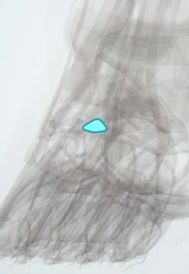
The spring ligament holds up the head of the talus from below like a sling; it is a key support of the inner arch of the foot.
Official name: Plantar calcaneonavicular ligament
Opens in the Durgical app, no sign-up needed.
What it is
A thick ligament under the inner midfoot, from the heel bone to the navicular. It cradles the head of the talus like a sling.
What it does
Holds up the head of the talus and supports the inner arch together with the tibialis posterior tendon.
Key facts
- Attaches
- Sustentaculum tali to plantar surface of navicular (plantar calcaneonavicular ligament)
- Function
- Supports head of talus; key support of medial longitudinal arch; tibialis posterior tendon supports it
- Tight in
- Weight-bearing (arch loaded); eversion
Relations
- Above
- Head of the talus, which rests on its fibrocartilage surface as part of the talocalcaneonavicular joint
- Medial
- Tibialis posterior tendon, which runs along its medial and plantar side on the way to the navicular tuberosity
- Lateral
- Calcaneonavicular limb of the bifurcate ligament; together they complete the socket for the talar head (acetabulum pedis)
- Below
- Flexor hallucis longus tendon under the sustentaculum tali and, more superficially, the abductor hallucis and medial plantar nerve
How to find it
Not palpable directly: it lies in the gap between the sustentaculum tali (a finger's breadth below the tip of the medial malleolus) and the navicular tuberosity, under the tibialis posterior tendon.
Examination
- Single-limb heel rise: inability to rise, or a heel that fails to swing into varus, suggests tibialis posterior and spring ligament failure
- 'Too many toes' sign: seen from behind, more lateral toes show on the affected side because the forefoot has drifted into abduction
On imaging
Standing lateral X-ray: the line through the talus and first metatarsal (Meary's angle), normally straight, sags toward the sole; standing AP: the talar head is uncovered by the navicular. MRI or ultrasound shows thinning, thickening or tears of the superomedial band.
Common problems
- Adult-acquired flatfoot: when the tibialis posterior tendon weakens, this ligament stretches or tears and the arch slowly collapses.
- Symptoms: pain and swelling on the inner ankle and arch, and the foot rolling inward.
How it is diagnosed
A physical exam, including standing on tiptoe on one foot (single heel rise). Standing X-rays; MRI or ultrasound.
Treatment
- Early stages: a brace or arch supports, and physical therapy to strengthen the tibialis posterior.
- Surgery: repair or reconstruction of the ligament, often with a tendon transfer and bone cuts to realign the foot. A graft from the patient or a donor may be used.
- Advanced, stiff flatfoot: fusion of the joints of the back of the foot.
Clinical notes
- As the support fails, the talus plantarflexes and turns inward, the heel drifts into valgus and the forefoot into abduction
- Spring ligament repair or reconstruction is often added to tendon transfer and calcaneal osteotomy in flexible flatfoot surgery
Related conditions
Test yourself
Think of the answer, then tap the question to see it.
What are the attachments of the spring (plantar calcaneonavicular) ligament?
From the sustentaculum tali to the plantar surface of the navicular.
What does the spring ligament support?
The head of the talus, which rests on its fibrocartilage surface; it is a key support of the medial longitudinal arch.
Which tendon works with the spring ligament, and what happens when that tendon fails?
The tibialis posterior; the ligament stretches, the talar head sags and a planovalgus (adult-acquired flat) foot develops.
What is the 'too many toes' sign?
Seen from behind, more lateral toes show on the affected side because the forefoot has drifted into abduction.
Educational information, not medical advice. For any symptom, see a health professional.
Its names
- Official Latin (TA2)
- ligamentum calcaneonaviculare plantare TA2 1937
- Official Spanish (DeCS)
- Ligamento calcaneonavicular plantar
- English (TA2)
- Plantar calcaneonavicular ligament
- Also called
- ligamentum calcaneonaviculare
Related
Sources: Terminologia Anatomica (TA2); Anatomy, Bony Pelvis and Lower Limb: Medial Longitudinal Arch of the Foot; Anatomy, Bony Pelvis and Lower Limb, Foot Joints.
Spotted a mistake?We are always improving for the community. Tell us what is wrong and our team will review it.Report a mistake
Representative model, for educational purposes only.