Cardiovascular system · Head and neck
Posterior communicating artery !
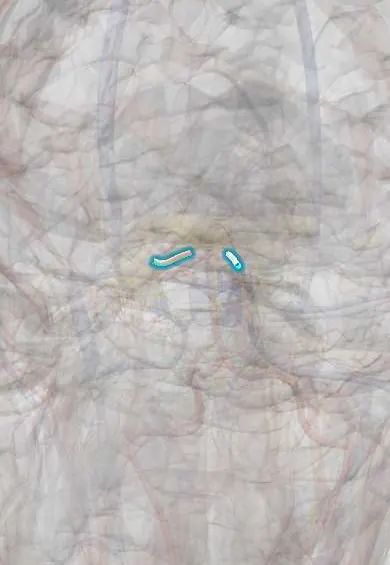
The posterior communicating artery links the front and back blood supply of the brain; an aneurysm here can press on the nerve that lifts the eyelid.
Opens in the Durgical app, no sign-up needed.
What it is
A short artery joining the internal carotid to the posterior cerebral artery; a common aneurysm site that can press on nerve III.
What it does
Links front and back circulations; an aneurysm here can press on nerve III (droopy lid, wide pupil).
Key facts
- Origin
- Internal carotid
- Branches
- Small perforators to the thalamus and hypothalamus
- Supplies
- Links carotid and vertebrobasilar systems; aneurysm here compresses CN III
Relations
- Course
- Leaves the back of the internal carotid just above the cavernous sinus and runs back, above the oculomotor nerve, to join the posterior cerebral artery at the P1–P2 junction
- Below
- Oculomotor nerve, running forward below and lateral to it
- Medial
- Tuber cinereum, the pituitary stalk and the mammillary bodies
- Above
- Optic tract and the base of the hypothalamus
How to find it
It cannot be felt. It runs back from the internal carotid to the posterior cerebral artery, above and medial to the oculomotor nerve and below the optic tract; in a pterional craniotomy it is seen behind the carotid once the carotid is gently lifted.
Examination
- Third nerve examination: lid position, eye position and movements, and the size and light reaction of both pupils
On imaging
CT or MR angiography shows its size and any aneurysm; catheter angiography confirms. A small funnel-shaped widening at its origin (infundibulum) can be mistaken for an aneurysm.
Common problems
- Brain aneurysm: a weak bulge in the artery wall, most often at this junction.
- Rupture causes a subarachnoid haemorrhage: a sudden 'worst headache of my life'.
How it is diagnosed
CT and CT angiography; angiography with a catheter to plan treatment; unruptured aneurysms are often found by chance on scans.
Treatment
- Clipping: a small metal clip across the neck of the aneurysm through a craniotomy.
- Coiling: soft platinum coils packed inside it through a catheter from the groin or wrist.
- Watching some small unruptured aneurysms, with blood pressure control and no smoking.
Clinical notes
- With a fetal-type posterior communicating artery, emboli from carotid disease can reach the occipital lobe, so an occipital stroke does not always mean a vertebrobasilar source
- A small funnel-shaped widening at its origin (infundibulum) is common and harmless, but on angiography it can be mistaken for an aneurysm
- Small perforating branches leave it for the thalamus, hypothalamus and internal capsule, so clipping or coiling of an aneurysm here must keep the artery open
Anatomical variants
- Often small (hypoplastic) on one or both sides
- Fetal pattern: a large posterior communicating artery is the main supply of the posterior cerebral artery
Development
It starts as the main caudal branch of the internal carotid, which first supplies the territory of the future posterior cerebral artery; the vertebrobasilar system later takes most of that flow, and when it does not, a fetal-type posterior communicating artery remains.
Memory aid
Rule of the pupil: pupil involved, think pressure (aneurysm); pupil spared, think small vessels (diabetes, hypertension).
Related conditions
Test yourself
Think of the answer, then tap the question to see it.
Which cranial nerve does a posterior communicating artery aneurysm compress, and what are the signs?
The oculomotor nerve (III): ptosis, eye down and out, and a dilated, unreactive pupil.
Why is a painful third nerve palsy with a dilated pupil treated as an emergency?
It suggests a posterior communicating artery aneurysm until imaging rules it out; microvascular palsies usually spare the pupil.
Which arteries does the posterior communicating artery join?
The internal carotid to the posterior cerebral artery (P1–P2 junction), linking the carotid and vertebrobasilar systems.
Educational information, not medical advice. For any symptom, see a health professional.
Its names
- Official Latin (TA2)
- arteria communicans posterior TA2 4521
- Official Spanish (DeCS)
- Arteria comunicante posterior
- English (TA2)
- Posterior communicating artery
Related
Sources: Terminologia Anatomica (TA2); Neuroanatomy, Circle of Willis; Anatomy of the Human Body (20th ed.): Development of the Vascular System (Lea & Febiger (Gray's Anatomy, 1918), 1918) · adapted, Public domain.
Spotted a mistake?We are always improving for the community. Tell us what is wrong and our team will review it.Report a mistake
Representative model, for educational purposes only.