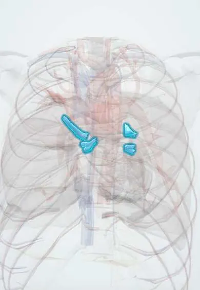
Pulmonary veins !
The pulmonary veins, usually four, carry oxygen-rich blood from the lungs back to the left atrium of the heart.
Opens in the Durgical app, no sign-up needed.
What it is
Four veins (two from each lung) bringing oxygen-rich blood back to the left atrium.
What it does
Return blood from the lungs to the heart. Extra electrical impulses from their openings often trigger atrial fibrillation.
Key facts
- Drains
- Lungs (oxygenated blood); usually superior and inferior vein per lung; right middle lobe vein joins right superior
- Into
- Left atrium, four valveless openings (varies: common left trunk)
Relations
- Course
- They begin in the intersegmental planes, join into lobar veins and usually leave each hilum as two veins, which pierce the pericardium and enter the back of the left atrium
- In front
- Right: the superior vena cava and right atrium lie in front of the right pulmonary veins as they cross to the left atrium
- Behind
- The oblique pericardial sinus, bounded by the four veins behind the left atrium; then the esophagus and, on the left, the descending thoracic aorta
- Below
- The pulmonary ligament, below the inferior pulmonary vein
How to find it
Not palpable. When the pericardium is opened and the heart lifted, the four veins are seen entering the back of the left atrium around the oblique pericardial sinus. At the hilum the inferior vein is reached by dividing the pulmonary ligament, a standard step in a lower lobectomy.
On imaging
CT and MRI map the number and openings of the veins before ablation. On a chest X-ray, raised left atrial pressure makes the upper lobe vessels wider than the lower ones (upper lobe diversion), an early sign of congestion.
Common problems
- Triggers of atrial fibrillation.
Treatment
- Catheter ablation isolates the pulmonary veins electrically to treat atrial fibrillation.
Clinical notes
- The esophagus lies just behind the left atrium, so ablation near the posterior wall can injure it; an atrioesophageal fistula is a rare, serious complication
- Narrowing of a pulmonary vein can follow ablation and cause breathlessness or haemoptysis
- In anomalous pulmonary venous return some or all veins drain to the right side, often with a sinus venosus atrial septal defect
Anatomical variants
- A common left trunk with a single opening and an extra right middle lobe vein are the commonest variants; four separate openings remain the usual pattern
- Partial anomalous pulmonary venous return, often a right upper vein draining into the superior vena cava
Development
A single common pulmonary vein buds from the back of the primitive left atrium and is absorbed into its wall; this forms the smooth part of the left atrium and leaves four veins opening separately.
Related conditions
- Truncus arteriosus, TAPVR and tricuspid atresia
- Heart failure
- Atrial fibrillation
- Rheumatic heart disease
- Mitral regurgitation
Test yourself
Think of the answer, then tap the question to see it.
Why does raised left atrial pressure cause pulmonary oedema?
The pulmonary veins have no valves, so pressure from mitral stenosis or left heart failure passes straight back to the lung capillaries.
Why are the pulmonary veins the target of catheter ablation for atrial fibrillation?
Sleeves of atrial muscle extend into their openings and fire ectopic beats; ablation isolates the veins electrically.
Which structure lying behind the left atrium can be injured during pulmonary vein ablation?
The esophagus; an atrioesophageal fistula is a rare, serious complication.
Where are the pulmonary veins at the hilum, and how is the inferior pulmonary vein reached in lower lobectomy?
They are the most anterior and lowest structures; the inferior vein is reached by dividing the pulmonary ligament below it.
Educational information, not medical advice. For any symptom, see a health professional.
Its names
- Official Latin (TA2)
- venae pulmonales TA2 4107
- Official Spanish (DeCS)
- Venas pulmonares DeCS D011667
- English (TA2)
- Pulmonary veins
Related
Sources: Terminologia Anatomica (TA2); DeCS; Anatomy, Thorax, Lung Veins; Anatomy, Thorax, Lungs.
Spotted a mistake?We are always improving for the community. Tell us what is wrong and our team will review it.Report a mistake
Representative model, for educational purposes only.