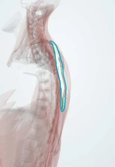
Rhomboids !
The rhomboids are muscles between the upper thoracic spine and the inner edge of the shoulder blade; they pull the shoulder blades together.
Official name: Rhomboid major muscle
Opens in the Durgical app, no sign-up needed.
What it is
Muscles between the upper thoracic spinous processes and the inner edge of the shoulder blade.
What it does
Pull the shoulder blades together.
Key facts
- Origin
- Minor: nuchal ligament and spinous processes of C7–T1; major: spinous processes of T2–T5
- Insertion
- Medial border of scapula: minor at root of the spine, major from spine to inferior angle
- Action
- Retract the scapula; rotate glenoid inferiorly; fix scapula to the thoracic wall
- Nerve
- Dorsal scapular nerve (C5; some texts: C4–C5)
- Blood supply
- Dorsal scapular artery (or deep branch of transverse cervical artery); posterior intercostal arteries
Relations
- Superficial
- Trapezius
- Deep
- Serratus posterior superior, splenius and the erector spinae; the dorsal scapular nerve and artery along the scapular border
- Above
- Levator scapulae, continuing the same line of insertion higher on the medial border
- Lateral
- Medial border of the scapula; at its lower edge, rhomboid major borders the triangle of auscultation
How to find it
With the hand on the hip and the elbow pushed backward against resistance, the rhomboids are felt under trapezius between the thoracic spine and the medial border of the scapula.
Examination
- Rhomboid test: hand on hip, the person pushes the elbow back against the examiner's hand; weakness with a medial border that lifts from the chest points to the dorsal scapular nerve or the C5 root
On imaging
Rarely imaged on their own; axial MRI or CT shows them deep to trapezius, between the thoracic spinous processes and the medial border of the scapula. Ultrasound shows them over the ribs, with the dorsal scapular artery and nerve on their deep side, for blocks and injections. In pain or weakness at the medial scapular border, imaging looks at the C5 root, scapula and chest wall.
Clinical notes
- The dorsal scapular nerve can be trapped as it passes through the middle scalene, giving aching along the medial border of the scapula
- Triangle of auscultation: with the arms folded and the trunk bent forward, this gap with little muscle over the 6th intercostal space lets breath sounds be heard clearly
Anatomical variants
- Rhomboid major and minor are often fused into one sheet with no clear gap between them; the number of spinous processes they arise from varies
Development
They form from tissue in the lower neck and migrate down to the medial border of the scapula, bringing the dorsal scapular nerve (C5) with them.
Test yourself
Think of the answer, then tap the question to see it.
What nerve supplies the rhomboids, and why does it localise a lesion?
The dorsal scapular nerve, which leaves the C5 root directly, so rhomboid weakness places the lesion at the root, not the trunks.
What are the attachments of the rhomboid major and minor?
Minor: C7–T1 spines to the root of the scapular spine; major: T2–T5 spines to the medial border down to the inferior angle.
Which triangle do the rhomboid major, trapezius and latissimus dorsi bound?
The triangle of auscultation, where breath sounds are heard clearly with the arms folded and trunk bent forward.
Educational information, not medical advice. For any symptom, see a health professional.
Its names
- Official Latin (TA2)
- musculus rhomboideus major; musculus rhomboideus minor TA2 2232
- Official Spanish (DeCS)
- Músculos romboides DeCS D064171
- English (TA2)
- Rhomboid major muscle
Related
Sources: Terminologia Anatomica (TA2); DeCS; Anatomy, Back, Extrinsic Muscles; Anatomy of the Human Body (20th ed.): Development of the Muscles (Lea & Febiger (Gray's Anatomy, 1918), 1918) · adapted, Public domain.
Spotted a mistake?We are always improving for the community. Tell us what is wrong and our team will review it.Report a mistake
Representative model, for educational purposes only.