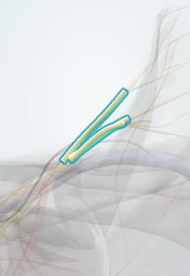
Plexus trunks !
The brachial plexus trunks are three nerve bundles (upper, middle, lower) in the base of the neck; they mix the spinal nerve roots on their way to the arm.
Official name: Brachial plexus trunks
Opens in the Durgical app, no sign-up needed.
What it is
Three thick bundles formed as the roots join, above the collarbone.
What it does
Mix the roots so each arm nerve gets fibres from several levels.
Key facts
- Roots
- Superior C5–C6, middle C7, inferior C8–T1
- Moves
- Superior trunk: suprascapular nerve, nerve to subclavius
- Feeling from
- Via branches (Erb: upper trunk; Klumpke: lower trunk)
Relations
- Course
- The roots leave the gap between the anterior and middle scalene muscles and join near the lateral border of the scalenes; the trunks run down and laterally across the posterior triangle of the neck toward the back of the clavicle, where each splits into divisions
- In front
- Platysma and deep cervical fascia; crossed by the inferior belly of the omohyoid and the transverse cervical and suprascapular vessels
- Medial
- Anterior scalene, with the phrenic nerve on its front; the subclavian artery lies in front of and below the lower trunk
- Behind
- Middle scalene
- Below
- The first rib, on which the lower trunk lies
How to find it
Above the clavicle, in the angle between the posterior border of the sternocleidomastoid and the clavicle, the trunks can sometimes be felt as firm cords that roll under the fingertip, more easily with the head turned to the other side.
Examination
- Adson test: neck extended and turned toward the tested side with a deep breath held; a fading radial pulse with reproduced symptoms suggests thoracic outlet compression, though false positives are common
- Elevated arm stress test (Roos): arms in 90° abduction and external rotation while opening and closing the hands for 3 minutes; early heaviness, pain or tingling supports thoracic outlet syndrome
- Pattern test: weak shoulder abduction, external rotation and elbow flexion with a lost biceps reflex points to the upper trunk; weak finger flexion and intrinsics with medial forearm numbness points to the lower trunk
On imaging
Ultrasound above the clavicle shows the trunks and divisions as a cluster of dark round bundles ('bunch of grapes') lateral and posterior to the subclavian artery, over the first rib. MRI with fat-suppressed fluid-sensitive sequences shows swelling and signal change after traction or in neuritis.
Common problems
- Stretch injuries from motorcycle crashes or falls that force the head and shoulder apart.
- Birth injury (Erb's palsy) affecting the upper trunk: the arm hangs turned inward ('waiter's tip').
- Compression at the thoracic outlet between the collarbone and the first rib.
How it is diagnosed
A detailed exam of strength and feeling in each nerve's area. MRI, EMG and nerve conduction studies show the level and severity.
Treatment
- Physical therapy and watching for recovery; many stretch injuries improve.
- Nerve repair, graft or nerve transfer surgery for injuries that do not recover, ideally within months.
Clinical notes
- Upper trunk injury comes from forced widening of the neck–shoulder angle: shoulder dystocia at birth, falls onto the shoulder from a motorcycle
- Lower trunk injury comes from forced arm abduction (breech delivery with the arm overhead, grabbing to stop a fall): claw hand from loss of the intrinsics
- Neurogenic thoracic outlet syndrome usually involves the lower trunk stretched over a cervical rib or fibrous band; long-standing cases waste the thenar eminence most (Gilliatt–Sumner hand)
- A Pancoast tumour at the lung apex invades the lower trunk and the sympathetic chain: shoulder and medial arm pain, hand weakness and Horner syndrome
- Interscalene and supraclavicular blocks are placed at root and trunk level; the phrenic nerve on the anterior scalene is often blocked as well, paralysing that hemidiaphragm
Anatomical variants
- Prefixed plexus: a large C4 contribution and a small T1; postfixed plexus: a small C5 and a T2 contribution; either shifts the root levels expected from a given lesion
- A cervical rib or a fibrous band from C7 can lift and kink the lower trunk
Development
The nerves reach the limb bud while it still sits opposite the lower neck; as the limb comes to lie lower on the trunk the roots slope downward, so the lower trunk arches over the first rib, where a cervical rib (an enlarged C7 costal element) can lift and kink it.
Memory aid
Roots, Trunks, Divisions, Cords, Branches: 'Randy Travis Drinks Cold Beer'.
Related conditions
Test yourself
Think of the answer, then tap the question to see it.
Which roots form the upper, middle and lower trunks of the brachial plexus?
Upper C5–C6, middle C7 and lower C8–T1; they cross the posterior triangle of the neck, with the lower trunk on the first rib.
What arm posture follows an upper trunk (Erb) palsy of the brachial plexus?
The arm hangs adducted and internally rotated, elbow straight, forearm pronated: deltoid, supra- and infraspinatus, biceps, brachialis and brachioradialis are lost.
What does a lower trunk (Klumpke) palsy of the brachial plexus cause, and what does Horner syndrome add?
Weak long finger flexors and all small hand muscles (claw hand) with medial forearm numbness; Horner syndrome adds a T1 root injury.
Which brachial plexus trunk is usually involved in neurogenic thoracic outlet syndrome, and over what?
The lower trunk, stretched over a cervical rib or a fibrous band from C7; long-standing cases waste the thenar eminence most (Gilliatt–Sumner hand).
Why can an interscalene block paralyse a hemidiaphragm?
The phrenic nerve lies on the front of the anterior scalene, next to the plexus roots and trunks, and is often blocked as well.
Educational information, not medical advice. For any symptom, see a health professional.
Its names
- Official Latin (TA2)
- truncus superior plexus brachialis TA2 6398
- Official Spanish (DeCS)
- Troncos superior, medio e inferior del plexo braquial
- English (TA2)
- Brachial plexus trunks
- Also called
- upper, middle and lower trunks
Related
Sources: Terminologia Anatomica (TA2); Anatomy, Shoulder and Upper Limb, Brachial Plexus; Anatomy, Head and Neck: Brachial Plexus; Anatomy of the Human Body (20th ed.): Development of the Muscles (Lea & Febiger (Gray's Anatomy, 1918), 1918) · adapted, Public domain.
Spotted a mistake?We are always improving for the community. Tell us what is wrong and our team will review it.Report a mistake
Representative model, for educational purposes only.