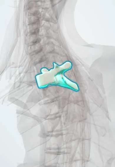
T2 vertebra !
T2 is the second thoracic (mid-back) vertebra; it has joints for the ribs and helps form the cage that protects the heart and lungs.
Official name: T2
Opens in the Durgical app, no sign-up needed.
What it is
The T2 mid-back vertebra, with joints on each side for the ribs.
What it does
Holds up the trunk and, with the ribs and breastbone, forms the rib cage, which protects the heart and lungs. The ribs make this part of the spine stiff.
Key facts
- Joints with
- T1 above and T3 below (discs, facet joints); heads of ribs 2 and 3; tubercle of rib 2 (costotransverse joint)
- Landmarks
- Typical thoracic: heart-shaped body; costal demifacets above and below; transverse costal facets; long sloping spine; jugular notch at about T2–T3 level
Relations
- In front
- Esophagus with the trachea in front of it; the left recurrent laryngeal nerve climbs in the groove between them; the brachiocephalic vessels lie farther forward
- Lateral
- Heads of the second and third ribs with the sympathetic trunk lying on them; the superior intercostal artery descends over the necks of the upper ribs
- Behind
- Trapezius, rhomboids (major, from T2 down) and serratus posterior (superior) over the erector spinae
- Contents
- Spinal cord, whose segments run about two levels below the vertebral number in the upper thoracic spine
How to find it
With the arms relaxed by the sides, a line joining the superior angles of the two scapulae crosses the T2 spine; counting down from C7 and T1 confirms it. In front, the jugular notch of the sternum lies at about the same height.
On imaging
The shoulders hide the upper thoracic spine on lateral X-rays, so fractures and lesions here are best seen on CT or MRI; sagittal whole-spine MRI allows counting down from C2.
Common problems
- Compression fractures from osteoporosis, sometimes after a minor fall or even a cough.
- A rounded upper back (kyphosis) from posture, wedge fractures or Scheuermann disease in teens.
- Scoliosis: a sideways curve with rotation, often starting in adolescence.
How it is diagnosed
X-rays for fractures and curves (scoliosis is measured with the Cobb angle). MRI tells new fractures from old ones. A bone density scan (DEXA) checks for osteoporosis.
Treatment
- Osteoporosis fractures: pain control, a short time in a brace, and treatment of the osteoporosis.
- Painful fractures that do not settle: kyphoplasty or vertebroplasty (cement injected into the bone).
- Large or progressive curves: bracing in growing children, sometimes fusion surgery.
Clinical notes
- The thoracic spine is the commonest site of metastatic spinal cord compression; upper thoracic deposits are easily missed on plain films
- Upper thoracic pain with stiffness in a young adult can be ankylosing spondylitis; a fracture through a fused spine after minor trauma is very unstable
- A cord injury at this level cuts the sympathetic outflow below it off from brain control, so autonomic dysreflexia can follow in the chronic phase
- The intercostobrachial nerve (from T2) supplies the armpit and inner upper arm; it is often stretched or cut in axillary node surgery, leaving numbness or pain there
Development
The thoracic spine keeps the forward-bent (kyphotic) curve of the fetus: it is a primary curve, while the neck and lower back develop their opposite curves after birth.
Test yourself
Think of the answer, then tap the question to see it.
Which scapular landmark lies opposite the spine of the T2 vertebra with the arms by the sides?
The superior angle of the scapula; the jugular notch of the sternum lies at about the same height in front.
Which artery supplies the first two intercostal spaces, at the level of the T2 vertebra?
The superior intercostal artery, from the costocervical trunk, not the aorta.
Why can autonomic dysreflexia follow a chronic spinal cord injury at the T2 vertebra?
The injury cuts the sympathetic outflow below it off from brain control.
Educational information, not medical advice. For any symptom, see a health professional.
Its names
- Official Latin (TA2)
- vertebra thoracica II TA2 1059
- Official Spanish (DeCS)
- Vértebra dorsal D2 DeCS D013904
- English (TA2)
- T2
Related
- T1–T2 disc
- T2–T3 disc
- Posterior intercostal arteries
- Anterior longitudinal ligament
- Rib-to-spine ligaments
- Dura (dural sac)
- Endplates
- Mid-back facet joints
Sources: Terminologia Anatomica (TA2); DeCS; Anatomy, Back, Thoracic Vertebrae; 7.3 The Vertebral Column (OpenStax (Rice University), 2022) · adapted, CC BY 4.0.
Spotted a mistake?We are always improving for the community. Tell us what is wrong and our team will review it.Report a mistake
Representative model, for educational purposes only.