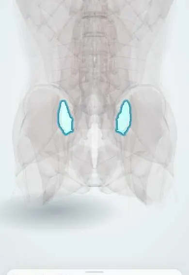
Ankylosing spondylitis !
Ankylosing spondylitis is a long-term inflammatory arthritis of the spine and sacroiliac joints; it causes back pain and stiffness and can fuse the spine.
Updated 2026-10-29
Structures involved
Tap one to see what it is and open it in 3D.
- SI joint surfaces
- Anterior longitudinal ligament
- Endplates
- Low-back facet joints
- Sacrum
- Sacroiliac ligaments
- Rib-to-spine ligaments
- Ribs (back part)
- T12 vertebra
- L1 vertebra
In immunology
In the clinical exam
Why it happens
- Chronic inflammation: causes, cells and features
- Synovial joints: capsule, synovial membrane and articular cartilage
- Ossification, growth plate and bone remodeling
What it is
Chronic inflammatory arthritis of the spine and sacroiliac joints, part of the spondyloarthritis family, that can fuse the spine. Today it is often called radiographic axial spondyloarthritis.
Who gets it
Usually starts in late adolescence or early adulthood, before age 45. The radiographic form is more common in men. Diagnosis is often delayed by years because early back pain is taken for mechanical pain.
Causes
- Immune-mediated inflammation of entheses (where ligaments and tendons attach to bone)
- Strong genetic link with HLA-B27
- Gut and other microbial triggers are thought to contribute
Risk factors
- HLA-B27 positive
- Family history of spondyloarthritis
- Male sex
- Associated psoriasis, inflammatory bowel disease or anterior uveitis
- Smoking (linked to more severe structural damage)
Pathophysiology
Inflammation starts at entheses and in the sacroiliac joints, with bone marrow oedema and erosions. Healing produces new bone: syndesmophytes bridge the vertebrae along the outer annulus and longitudinal ligaments, and the facet and sacroiliac joints fuse. Full fusion gives the bamboo spine; the rigid, osteoporotic spine breaks easily.
Classification
- Radiographic axial spondyloarthritis (ankylosing spondylitis): sacroiliitis visible on X-ray
- Non-radiographic axial spondyloarthritis: inflammation on MRI or clinical features without X-ray changes
- Classification criteria (ASAS; modified New York for the radiographic form) are for research, not for diagnosing a single patient
Symptoms and signs
- Inflammatory back pain: slow onset, lasting more than three months, worse with rest and better with exercise
- Morning stiffness lasting more than about 30 minutes; night pain that wakes the patient in the second half of the night
- Alternating buttock pain from sacroiliitis
- Progressive loss of spinal movement and stooped posture (kyphosis)
- Enthesitis (heel or Achilles pain), peripheral arthritis or dactylitis
- Anterior uveitis: painful red eye with light sensitivity (the most common extra-articular feature)
- Reduced chest expansion and fatigue
Diagnosis
- Clinical
- Inflammatory back pain in a young adult plus exam: modified Schober test (classically less than 5 cm of increase on forward bending), reduced chest expansion, occiput-to-wall distance and lumbar side flexion. Activity scores such as BASDAI and ASDAS follow the disease.
- Tests
- CRP and ESR may be raised but are often normal. HLA-B27 supports the diagnosis but is neither necessary nor sufficient. Rheumatoid factor is negative.
- Imaging
- Pelvic X-ray shows sacroiliitis (sclerosis, erosions, fusion); spine X-ray shows syndesmophytes, squaring of vertebrae and bamboo spine. MRI of the sacroiliac joints detects early inflammation (bone marrow oedema) before X-ray changes.
Differential diagnosis
- Mechanical low back pain
- Diffuse idiopathic skeletal hyperostosis (DISH)
- Psoriatic arthritis or arthritis of inflammatory bowel disease
- Infectious sacroiliitis, including brucellosis or tuberculosis
- Osteitis condensans ilii
Goal of treatment
Control pain, stiffness and inflammation, keep posture and spinal mobility, prevent structural damage and treat the extra-articular disease.
Treatment
- Rehabilitation
- Regular exercise and physiotherapy for posture, spinal mobility and breathing are a cornerstone at every stage.
- Lifestyle
- Smoking cessation and education; attention to fracture risk and bone health.
- Medical
- NSAIDs are the first-line drug class and are continued when they control symptoms. Systemic glucocorticoids are not used for axial disease; conventional synthetic DMARDs help peripheral arthritis only.
- Medical
- Biologic therapy, TNF inhibitors or IL-17 inhibitors, when disease stays active despite NSAIDs; JAK inhibitors are another option. TB is screened for first, an important step in Colombia.
- Surgical
- Surgery for selected patients: hip replacement for destroyed hips, corrective osteotomy for severe fixed kyphosis, and stabilisation of spinal fractures.
Drugs for this condition
- Celecoxib
- Indometacin
- Diclofenac
- Naproxen
- Adalimumab
- Infliximab
- Etanercept
- Certolizumab pegol
- Golimumab
- Secukinumab
- Tofacitinib
Complications
- Spinal fracture after minor trauma, often unstable and with cord injury
- Osteoporosis
- Fixed kyphotic deformity
- Recurrent anterior uveitis
- Aortic regurgitation and heart conduction disease
- Restrictive lung disease and apical lung fibrosis
Prognosis
A lifelong disease of varying course. Many patients keep a good working life; early diagnosis, exercise and modern drugs reduce symptoms, and biologics improve function and quality of life. Male sex, smoking, high CRP and existing syndesmophytes predict faster progression.
Test yourself
Think of the answer, then tap the question to see it.
Which features distinguish inflammatory back pain from mechanical back pain?
Onset before 45, insidious onset, improvement with exercise, no improvement with rest, and night pain, often with long morning stiffness.
Which HLA allele is strongly associated with ankylosing spondylitis?
HLA-B27. It supports the diagnosis but is not enough on its own, since many healthy people carry it.
What does the modified Schober test measure?
Lumbar flexion: the increase in distance between skin marks over the lumbar spine on bending forward. A small increase (classically under 5 cm) suggests restriction.
What is the most common extra-articular manifestation of ankylosing spondylitis?
Acute anterior uveitis: a painful red eye with photophobia.
What is the first-line drug class for axial spondyloarthritis?
NSAIDs. If disease remains active, biologics such as TNF or IL-17 inhibitors are added.
Related conditions
- Sacroiliac joint pain
- Mechanical low back pain
- Osteoporotic vertebral compression fracture
- Spinal cord injury
- Rheumatoid arthritis
- Crohn disease
- Ulcerative colitis
- Uveitis
Educational information, not medical advice. For any symptom, see a health professional.
Sources: Ankylosing Spondylitis (StatPearls (NCBI Bookshelf), 2023).
Spotted a mistake?We are always improving for the community. Tell us what is wrong and our team will review it.Report a mistake
Representative model, for educational purposes only.