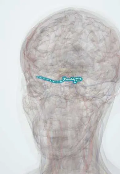
Nervous system · Head and neck
Trochlear nerve (IV) !
The trochlear nerve (cranial nerve IV) is the thinnest cranial nerve; it moves one eye muscle, and damage causes double vision when looking down.
Official name: Trochlear nerve
Opens in the Durgical app, no sign-up needed.
What it is
The thinnest cranial nerve, the only one leaving the back of the brainstem.
What it does
Supplies the superior oblique; a palsy gives double vision walking downstairs.
Key facts
- Roots
- Trochlear nucleus (midbrain); only nerve exiting the dorsal brainstem; superior orbital fissure
- Moves
- Superior oblique
- Feeling from
- None
Relations
- Course
- Exits the back of the midbrain, curves forward around the cerebral peduncle between the posterior cerebral and superior cerebellar arteries, pierces the dura at the edge of the tentorium, runs in the lateral wall of the cavernous sinus and enters the orbit through the superior orbital fissure, outside the common tendinous ring, crossing above the levator palpebrae to the superior oblique
- Above
- In the lateral wall of the cavernous sinus, the oculomotor nerve (III)
- Below
- In the lateral wall of the cavernous sinus, the V1 ophthalmic nerve
How to find it
It cannot be felt; it is assessed by having the person look down while the eye is turned in. In dissection it is a very thin thread that appears just below the inferior colliculus and winds around the midbrain at the edge of the tentorium.
Examination
- Eye movements: with the eye turned in, weak downward movement points to the superior oblique (CN IV)
- Three-step (Parks–Bielschowsky) test: a higher eye that gets worse on gaze to the opposite side and on head tilt toward the affected side points to a superior oblique palsy
- Head posture: a compensatory tilt away from the affected side; the same tilt in old photographs suggests a long-standing (congenital) palsy
On imaging
Very thin: only high-resolution heavily T2-weighted MRI shows its cisternal part winding around the midbrain in the ambient cistern. In a palsy, MRI checks the back of the midbrain below the inferior colliculus, the cavernous sinus and the orbit; after head injury, CT looks for associated damage. A small superior oblique on coronal orbital MRI supports a long-standing palsy.
Clinical notes
- Closed head injury is a classic cause: the nerve is stretched against the edge of the tentorium, and severe trauma can injure both sides
- A lesion of the nucleus weakens the opposite eye, because the fibres cross before they leave the brainstem
- Lesions of the cavernous sinus or the superior orbital fissure usually affect it together with III, V1 and VI
Memory aid
"RAD SIN": the recti adduct (except the lateral rectus), and the superior muscles (superior oblique, superior rectus) intort.
Test yourself
Think of the answer, then tap the question to see it.
What two features make the trochlear nerve (IV) unique among cranial nerves?
It is the only one leaving the back of the brainstem, and its fibres cross, so each nucleus supplies the opposite superior oblique.
When is double vision worst in a trochlear nerve palsy, and which way does the head tilt?
Looking down and in (reading, going downstairs); the head tilts away from the affected side to make up for lost intorsion.
Why is the trochlear nerve often injured in closed head trauma?
It is the thinnest cranial nerve with the longest course inside the skull, so it is stretched against the edge of the tentorium.
Which named test confirms a superior oblique (trochlear nerve) palsy?
The three-step Parks–Bielschowsky test: a higher eye worse on gaze to the opposite side and on head tilt toward the affected side.
Educational information, not medical advice. For any symptom, see a health professional.
Its names
- Official Latin (TA2)
- nervus trochlearis TA2 6191
- Official Spanish (DeCS)
- Nervio Troclear DeCS D014321
- English (TA2)
- Trochlear nerve
- Also called
- cranial nerve IV
Related
Sources: Terminologia Anatomica (TA2); DeCS; Neuroanatomy, Cranial Nerve 4 (Trochlear).
Spotted a mistake?We are always improving for the community. Tell us what is wrong and our team will review it.Report a mistake
Representative model, for educational purposes only.