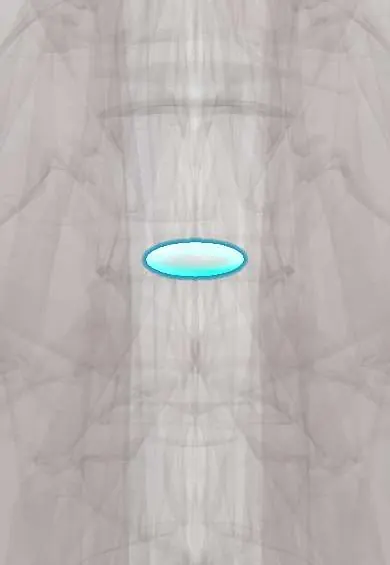
T11–T12 nucleus !
The nucleus pulposus is the soft, gel-like centre of the T11-T12 disc in the mid-back; it works like a water cushion that spreads the load.
Official name: T11-T12
Opens in the Durgical app, no sign-up needed.
What it is
The soft, gel-like centre (nucleus pulposus) of the T11–T12 disc.
What it does
Works like a water cushion: it turns pressure from above into outward push on the fibre ring, spreading the load. It is under the most pressure when you bend forward and lift.
Key facts
- Where
- Centre of the T11–T12 disc, slightly toward the back, inside the annulus fibrosus
- Function
- Notochord-derived gel (about 70–90% water, proteoglycans); turns axial load into annulus tension; small in thin thoracic discs, herniation rare
Relations
- Behind
- Back of the annulus fibrosus and the posterior longitudinal ligament, then the lower spinal cord near the conus medullaris
How to find it
Found on imaging, not by touch. The level is fixed by counting down from C2 on a whole-spine sagittal scout and checking it against the 12th rib, because wrong-level surgery is a known risk in the thoracic spine.
Examination
- Ankle jerks and toe strength, with perianal sensation and the anal reflex, test the lowest cord segments, which lie just behind and below this level
On imaging
Before surgery at this level, CT or MR angiography may be used to locate the artery of Adamkiewicz, which often enters the canal nearby.
Common problems
- Squeezes out through a tear in the ring (herniation), usually backward and to one side, toward a nerve root.
- Loses water with age, so the disc gets thinner and stiffer.
How it is diagnosed
On MRI a healthy nucleus is bright on T2 images; a worn one turns dark ('black disc').
Treatment
- In a microdiscectomy only the loose piece that has pushed out is removed; most of the disc stays.
Clinical notes
- Epiconus syndrome: compression just above the conus (roughly the L4–S2 segments, texts vary) gives foot and ankle weakness with mixed reflexes, sometimes with early bladder trouble
- Thoracic herniations are far less common than cervical or lumbar ones, and many are incidental MRI findings; symptomatic fragments are often calcified, which makes removal harder
- These herniations are removed from the side or the front (transpedicular, costotransversectomy, thoracoscopic or open transthoracic routes); a plain laminectomy would need the cord to be retracted and carries a high risk of worsening the deficit
Development
The nucleus is what is left of the notochord: the notochord vanishes from the growing vertebral bodies but survives between them, where it helps build each disc.
Test yourself
Think of the answer, then tap the question to see it.
Why is the T11–T12 nucleus one of the most frequently herniated thoracic nuclei?
It sits at the junction where the rib cage stops splinting the spine.
Which artery may be located with CT or MR angiography before surgery on the T11–T12 nucleus?
The artery of Adamkiewicz, which often enters the canal near this level.
Which part of the spinal cord lies behind the T11–T12 nucleus pulposus?
The lower cord near the conus medullaris, behind the annulus fibrosus and the posterior longitudinal ligament.
Educational information, not medical advice. For any symptom, see a health professional.
Its names
- Official Latin (TA2)
- nucleus pulposus T11-T12 TA2 1686
- Official Spanish (DeCS)
- Núcleo pulposo T11-T12 DeCS D000070614
- English (TA2)
- T11-T12
Related
Sources: Terminologia Anatomica (TA2); DeCS; Anatomy, Back, Nucleus Pulposus; Anatomy, Back, Intervertebral Discs; 7.3 The Vertebral Column (OpenStax (Rice University), 2022) · adapted, CC BY 4.0; 7.5 Embryonic Development of the Axial Skeleton (OpenStax (Rice University), 2022) · adapted, CC BY 4.0.
Spotted a mistake?We are always improving for the community. Tell us what is wrong and our team will review it.Report a mistake
Representative model, for educational purposes only.