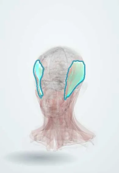
Myotonic dystrophy (types 1 and 2) !
An inherited muscle disease that causes weakness, slow relaxation of muscles after gripping, cataracts and heart rhythm problems.
Updated 2026-11-01
Structures involved
Tap one to see what it is and open it in 3D.
- Temporalis
- Sternocleidomastoid
- Masseter
- Levator palpebrae
- Eyelids
- Orbicularis oculi
- Lens
- Pharynx
- Atrioventricular (AV) node
- Bundle of His
- Bundle branches
- Diaphragm
- APB
- Dorsal interossei
- Tibialis anterior
Why it happens
- Mendelian inheritance: autosomal dominant, recessive and X-linked
- Nonclassic inheritance: repeat expansions, mitochondrial genes, imprinting, mosaicism
- Ion channels and gating
- Trinucleotide repeat expansion disorders
- RNA processing (cap, poly-A tail and splicing)
What it is
An autosomal dominant multisystem disorder in which muscles weaken and waste and are slow to relax after contraction (myotonia). It also affects the heart conduction system, eyes, endocrine glands, gut and brain. It is the most common muscular dystrophy that begins in adulthood. Type 1 (DM1, Steinert disease) is more frequent and more severe; type 2 (DM2, proximal myotonic myopathy) is milder.
Who gets it
Affects at least about 1 in 8,000 people worldwide. DM1 is the predominant type in most populations, including Latin America; DM2 is relatively frequent in northern and central Europe. Symptoms usually start in the twenties or thirties, but a severe congenital DM1 form presents at birth.
Causes
- DM1: expansion of a CTG trinucleotide repeat in the DMPK gene on chromosome 19
- DM2: expansion of a CCTG repeat in the CNBP gene on chromosome 3
- Autosomal dominant inheritance
- Anticipation in DM1: the repeat lengthens across generations, giving earlier onset and more severe disease; the congenital form is usually inherited from the mother
Risk factors
- Affected parent (50% risk for each child)
- Maternal transmission of DM1 (risk of the congenital form)
- Large repeat size
Pathophysiology
The expanded repeats are transcribed into RNA that is not translated but accumulates in the nucleus. This toxic RNA traps RNA-binding proteins (such as muscleblind-like proteins), which disturbs the splicing of many other genes. Mis-splicing of the muscle chloride channel causes myotonia, of the insulin receptor causes insulin resistance, and of cardiac proteins contributes to conduction disease. This RNA gain-of-function mechanism explains why so many organs are involved.
Classification
- DM1: congenital, childhood-onset, classic adult and mild late-onset forms
- DM2: adult onset, mainly proximal weakness and pain, no congenital form
Symptoms and signs
- Grip myotonia: difficulty letting go after a handshake or gripping an object, improving with repetition (warm-up)
- Percussion myotonia of the thenar muscles or tongue
- Distal weakness in DM1 (hands, foot drop) and weakness of neck flexors; proximal weakness in DM2
- Characteristic face: ptosis, wasting of the temporalis and masseter ("hatchet face"), frontal balding in men
- Difficulty swallowing and nasal speech
- Early posterior subcapsular cataracts with iridescent opacities
- Palpitations, fainting from conduction block or arrhythmias
- Daytime sleepiness, apathy and cognitive problems
- Testicular atrophy, insulin resistance or diabetes
- Congenital DM1: floppy newborn, poor sucking, respiratory failure, club feet, later intellectual disability
Diagnosis
- Clinical
- Suspected from the combination of myotonia, typical facial weakness, early cataracts and a family history. Myotonia can be shown by slow hand opening after gripping or by tapping the thenar eminence.
- Tests
- Genetic testing for the repeat expansion in DMPK or CNBP confirms the diagnosis. EMG shows myotonic discharges ("dive bomber" sound). CK is normal or mildly raised. ECG often shows a prolonged PR interval or bundle branch block; glucose, HbA1c and testosterone are checked.
- Imaging
- Echocardiography and Holter monitoring for heart disease; slit-lamp eye examination for cataracts. Muscle biopsy is rarely needed now.
Differential diagnosis
- Other muscular dystrophies (facioscapulohumeral, limb-girdle)
- Non-dystrophic myotonias (myotonia congenita, paramyotonia)
- Inclusion body myositis and inflammatory myopathies
- Myasthenia gravis (ptosis and facial weakness)
- Hereditary neuropathies (distal weakness)
- Spinal muscular atrophy and congenital myopathies in the floppy infant
Goal of treatment
There is no cure; care aims to prevent sudden cardiac death and respiratory complications, treat symptoms and keep the person independent.
Treatment
- Follow-up
- Annual ECG and periodic Holter and echocardiography; screening for diabetes, thyroid disease and cataracts; sleep and lung function studies. Nursing priority: careful monitoring after sedation or anaesthesia because of high sensitivity to these drugs.
- Procedure
- Pacemaker or implantable cardioverter-defibrillator for conduction block or ventricular arrhythmia; non-invasive ventilation for hypoventilation.
- Medical
- Sodium-channel blockers such as mexiletine for disabling myotonia (after cardiac assessment); treatment of diabetes and hypogonadism; stimulants for excessive daytime sleepiness in some patients.
- Surgical
- Cataract surgery; eyelid surgery for severe ptosis.
- Rehabilitation
- Physiotherapy, ankle-foot orthoses for foot drop, speech and swallowing therapy, occupational therapy and genetic counselling for the family.
Complications
- Sudden cardiac death from heart block or ventricular arrhythmia
- Respiratory failure and aspiration pneumonia
- Anaesthetic complications
- Diabetes and infertility
- Falls and loss of mobility
- Gallstones and bowel dysmotility
Prognosis
Classic adult DM1 progresses slowly; life expectancy is reduced, mainly by respiratory and cardiac causes. DM2 is milder with near-normal survival. Congenital DM1 carries a high neonatal mortality and lasting intellectual disability in survivors.
Prevention
- Genetic counselling for affected families
- Prenatal or preimplantation testing when the family wishes
- Regular cardiac screening and timely pacemaker or defibrillator implantation to prevent sudden death
Test yourself
Think of the answer, then tap the question to see it.
A man cannot release his grip after a handshake and has early cataracts and frontal balding. What is the diagnosis?
Myotonic dystrophy type 1.
What genetic mechanism causes DM1?
An expanded CTG trinucleotide repeat in the DMPK gene, with anticipation across generations.
Which cardiac problem must be screened for and why?
Conduction block and arrhythmias, because they cause sudden death; screening is with ECG and Holter.
From which parent is congenital DM1 usually inherited?
From the mother.
What does EMG show?
Myotonic discharges that wax and wane in frequency and amplitude ("dive bomber" sound).
Related conditions
- Atrioventricular (heart) block
- Cataract
- Type 2 diabetes mellitus
- Obstructive sleep apnoea
- Male hypogonadism
- Atrial flutter
- Aspiration pneumonia and pneumonitis
- Oropharyngeal dysphagia in older adults
- Inflammatory myopathies (dermatomyositis, polymyositis)
- Charcot-Marie-Tooth disease
- Spinal muscular atrophy
Educational information, not medical advice. For any symptom, see a health professional.
Sources: Myotonic dystrophy: MedlinePlus Genetics (MedlinePlus (U.S. National Library of Medicine), 2025).
Spotted a mistake?We are always improving for the community. Tell us what is wrong and our team will review it.Report a mistake
Representative model, for educational purposes only.