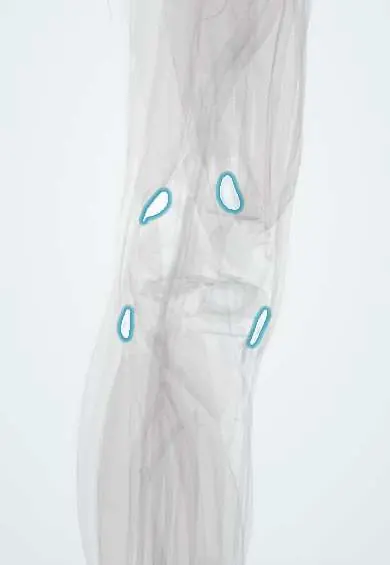
Back-of-knee bursae !
The back-of-knee bursae are small fluid sacs under the hamstring and calf tendons; one often links to the joint and forms Baker's cysts.
Official name: Semimembranosus bursa
Opens in the Durgical app, no sign-up needed.
What it is
Small fluid sacs under the hamstring and calf tendons at the back of the knee.
What it does
Reduce friction; a swollen one can form a Baker's cyst.
Key facts
- Where
- Semimembranosus and medial gastrocnemius bursae; lateral gastrocnemius bursa; popliteus recess
- Function
- Reduce tendon friction; gastrocnemius-semimembranosus bursa may swell as a popliteal (Baker) cyst
Relations
- In front
- Posterior capsule of the knee and the femoral condyles
- Behind
- Popliteal fascia and skin; the popliteal artery, popliteal vein and tibial nerve run down the middle of the fossa between the medial and lateral bursae
- Medial
- Semimembranosus bursa between that tendon and the medial tibial condyle; medial gastrocnemius bursa between the medial head and the capsule, often joined to it
- Lateral
- Lateral gastrocnemius bursa under the lateral head; popliteus recess between the popliteus tendon, the lateral meniscus and the lateral femoral condyle
How to find it
A Baker cyst is felt on the medial side of the popliteal fossa, between the medial head of gastrocnemius and the semimembranosus tendon. It is easiest to feel with the knee straight, when it becomes tense, and softens as the knee bends.
Examination
- Foucher sign: a popliteal cyst feels firm with the knee fully extended and softens at about 45° of flexion, which helps tell it from a solid mass or an aneurysm
On imaging
Ultrasound shows a fluid collection with a neck between the medial gastrocnemius and the semimembranosus tendon; a ruptured cyst leaves fluid tracking down the calf. Doppler excludes a popliteal artery aneurysm and deep vein thrombosis. MRI shows the cyst and the joint problem that fills it, such as a meniscal tear or osteoarthritis.
Clinical notes
- In adults most popliteal cysts are secondary to joint disease with a one-way valve, so they recur unless the joint problem settles; in children they are usually isolated and often regress
- Rupture causes sudden calf pain and swelling, sometimes with a crescent of bruising below the medial malleolus (crescent sign)
- A large cyst can compress the popliteal vein (leg swelling, thrombosis) or the tibial nerve
- A pulsatile popliteal mass is an aneurysm until shown otherwise, not a cyst
Anatomical variants
- Communication between the semimembranosus–gastrocnemius bursa and the knee joint is more frequent in older adults than in children, which matches the age pattern of popliteal cysts
Related conditions
Test yourself
Think of the answer, then tap the question to see it.
Which bursa at the back of the knee most often forms a Baker (popliteal) cyst, and why?
The semimembranosus–medial gastrocnemius bursa: it often connects with the knee joint through a slit in the posteromedial capsule, so joint fluid gets trapped in it.
What is the Foucher sign in a popliteal (Baker) cyst?
The cyst feels firm with the knee fully extended and softens at about 45° of flexion, which helps tell it from a solid mass or an aneurysm.
Which condition does a ruptured Baker cyst from the back-of-knee bursae mimic?
Deep vein thrombosis: the fluid tracks into the calf and causes sudden calf pain and swelling, sometimes with a crescent of bruising below the medial malleolus.
Educational information, not medical advice. For any symptom, see a health professional.
Its names
- Official Latin (TA2)
- bursa musculi semimembranosi; bursa subtendinea lateralis musculi gastrocnemii; bursa subtendinea medialis musculi gastrocnemii TA2 2748
- Official Spanish (DeCS)
- Bolsas del semimembranoso y subtendinosas del gastrocnemio DeCS D002061
- English (TA2)
- Semimembranosus bursa
- Also called
- bursa fibularis musculi semimembranacei · Brodie's bursa
Sources: Terminologia Anatomica (TA2); DeCS; Anatomy, Skin Bursa; Anatomy, Bony Pelvis and Lower Limb, Knee.
Spotted a mistake?We are always improving for the community. Tell us what is wrong and our team will review it.Report a mistake
Representative model, for educational purposes only.