Conditions · Ophthalmology and ENT
Cholesteatoma !
A growing pocket of skin trapped in the middle ear that slowly erodes bone. It causes smelly discharge and hearing loss and needs surgery.
Updated 2026-11-01
Structures involved
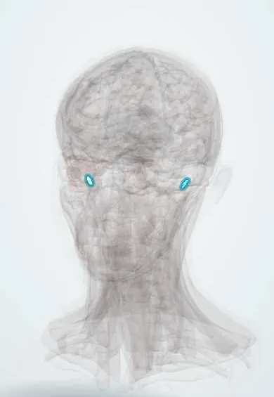
Tap one to see what it is and open it in 3D.
Why it happens
- Inner ear: the cochlea and the organ of Corti
- Simple, stratified, pseudostratified and transitional epithelium
What it is
A cyst-like mass of keratinising squamous epithelium and keratin debris in the middle ear and mastoid. It is benign but locally destructive, eroding the ossicles and surrounding bone.
Who gets it
Most cases are acquired and follow chronic otitis media or long-standing auditory tube dysfunction; congenital cases are rare and found in children behind an intact eardrum. It is a common reason for ear surgery where chronic ear disease is frequent, as in many parts of Latin America.
Causes
- Acquired, primary: a retraction pocket of the pars flaccida (attic) from chronic negative middle ear pressure that traps keratin
- Acquired, secondary: skin migrating into the middle ear through a marginal perforation
- Implantation after ear surgery or trauma
- Congenital: an embryonic rest of epithelium behind an intact eardrum
Risk factors
- Chronic or recurrent otitis media
- Auditory tube dysfunction, cleft palate and craniofacial anomalies
- Previous ear surgery or eardrum perforation
Pathophysiology
Trapped squamous epithelium keeps shedding keratin that cannot clear, so the sac enlarges. Enzymes and inflammation from the matrix and from infected debris resorb bone, destroying the ossicles (often the long process of the incus), the scutum and the bony covering of the lateral semicircular canal, facial nerve and tegmen. Anaerobic infection of the debris produces foul discharge.
Classification
- Congenital (intact eardrum, no history of ear infection)
- Acquired primary (attic retraction pocket)
- Acquired secondary (marginal or posterosuperior perforation)
Symptoms and signs
- Persistent or recurrent foul-smelling ear discharge that does not settle with drops
- Progressive one-sided conductive hearing loss
- Ear fullness or pressure
- On otoscopy: pearly white mass or keratin debris in an attic retraction pocket or marginal perforation, sometimes granulation tissue or a polyp
- Dizziness, especially with pressure on the ear, if the lateral semicircular canal is eroded (fistula)
Diagnosis
- Clinical
- Otomicroscopy after cleaning the canal shows the keratin mass or retraction pocket; the fistula test (vertigo or nystagmus with pressure in the canal) suggests labyrinthine erosion.
- Tests
- Audiometry documents the hearing loss before surgery; culture of discharge when infected.
- Imaging
- High-resolution CT of the temporal bone shows bone erosion and extent and plans surgery; diffusion-weighted MRI distinguishes cholesteatoma from inflammation and detects residual or recurrent disease.
Differential diagnosis
- Chronic suppurative otitis media without cholesteatoma
- Otitis externa or a foreign body
- Glomus tumour (paraganglioma) of the middle ear
- Middle ear or ear canal carcinoma
Goal of treatment
Remove all of the cholesteatoma to obtain a safe, dry ear, then preserve or rebuild hearing, and follow up long enough to detect recurrence.
Treatment
- Surgical
- Surgery is the definitive treatment: tympanomastoidectomy, either canal wall up (keeps the ear canal wall, often needs a second-look operation) or canal wall down (open cavity, lower recurrence), with ossicular reconstruction when possible.
- Medical
- Before surgery, cleaning of the ear and topical antibiotic drops reduce infection and discharge; they do not cure the disease.
- Follow-up
- Long-term follow-up with otomicroscopy and diffusion-weighted MRI to detect residual or recurrent disease; nursing care after surgery includes keeping the dressing dry and watching for facial weakness, vertigo and bleeding.
Complications
- Conductive and, with labyrinthine involvement, sensorineural hearing loss
- Labyrinthine fistula with vertigo
- Facial nerve palsy
- Mastoiditis and subperiosteal abscess
- Meningitis, epidural or brain abscess, and sigmoid sinus thrombosis
Prognosis
Surgery removes the disease in most patients, but recurrence or residual disease is common, especially in children, so many need repeated surgery and years of follow-up. Untreated, it keeps growing and can cause dangerous complications.
Prevention
- Timely treatment and follow-up of chronic ear infections and retraction pockets
Test yourself
Think of the answer, then tap the question to see it.
What is a cholesteatoma made of?
Keratinising squamous epithelium and accumulated keratin debris in the middle ear and mastoid.
Which otoscopic finding is classic for acquired cholesteatoma?
A pearly white mass or keratin in an attic (pars flaccida) retraction pocket or marginal perforation.
Which ossicle is most often eroded first?
The incus, especially its long process.
What is the definitive treatment of cholesteatoma?
Surgical removal (tympanomastoidectomy).
Related conditions
- Otitis media with effusion and chronic suppurative otitis media
- Acute mastoiditis
- Hearing loss (conductive and sensorineural, including sudden hearing loss)
- Bacterial meningitis
- Brain abscess
- Bell palsy
Educational information, not medical advice. For any symptom, see a health professional.
Sources: Cholesteatoma (MedlinePlus, US National Library of Medicine, 2026).
Spotted a mistake?We are always improving for the community. Tell us what is wrong and our team will review it.Report a mistake
Representative model, for educational purposes only.