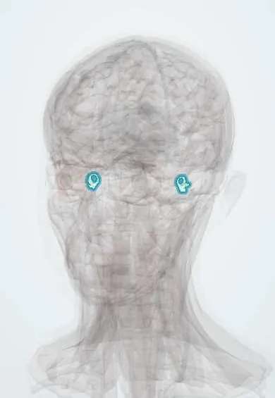
Vestibule !
The vestibule is the central chamber of the inner ear; it senses head tilt and straight-line movement, such as in a lift or a car.
Opens in the Durgical app, no sign-up needed.
What it is
The central chamber of the inner ear.
What it does
Senses head tilt and straight-line movement (gravity, lifts, cars).
Key facts
- Where
- Inner ear between the cochlea and the semicircular canals
- Blood supply
- Labyrinthine artery
- Nerve
- Vestibular part of VIII
- Function
- Utricle and saccule sense gravity and linear acceleration
Relations
- In front
- The cochlea, joined to the saccule by a fine duct
- Behind
- The three semicircular canals, which open into the vestibule through five openings
- Lateral
- The middle ear: the oval window with the stapes footplate, and the facial nerve running just above it
- Medial
- The fundus of the internal acoustic meatus, through which the vestibular nerves reach the utricle and saccule
- Contents
- Perilymph around the membranous utricle and saccule, which hold endolymph; the endolymphatic duct leaves through the vestibular aqueduct
How to find it
It can be neither seen nor felt. From the middle ear it lies deep to the oval window, above and behind the promontory, so an instrument passed through the stapes footplate in stapes surgery enters it; it sits between the cochlea in front and the semicircular canals behind.
Examination
- Bedside balance tests check it together with the semicircular canals: Romberg test, gait and the head-impulse test
- Positional tests such as the Dix–Hallpike manoeuvre look for loose otoconia
- Caloric testing (warm or cold water or air in the ear canal) checks each side's vestibular response by the eye movements it provokes
- Vestibular evoked myogenic potentials (VEMP) test the saccule and utricle in a hearing and balance unit
On imaging
High-resolution CT of the temporal bone shows the bony vestibule; MRI shows the fluid-filled labyrinth and the vestibular nerves, which matters when a vestibular schwannoma is suspected.
Clinical notes
- Benign paroxysmal positional vertigo starts when otoconia break loose from the utricle and drift into a semicircular canal, most often the posterior one
- Ménière disease is linked to too much endolymph in the membranous labyrinth: episodes of vertigo, hearing loss, tinnitus and ear fullness
- Vestibular neuritis inflames the vestibular nerve and gives days of severe vertigo without hearing loss
- A large vestibular aqueduct is one of the commonest inner-ear malformations seen on imaging in children with hearing loss
Development
The vestibule forms from the otic vesicle, which grows from a thickening of surface ectoderm (the otic placode) beside the hindbrain; the utricle gives rise to the semicircular ducts and the saccule to the cochlear duct.
Related conditions
Test yourself
Think of the answer, then tap the question to see it.
Which two otolith organs does the vestibule of the inner ear hold, and what do they sense?
The utricle (horizontal macula) and the saccule (vertical macula); calcium crystals on their hair cells make them sense gravity and straight-line acceleration.
What sits in the oval window on the lateral wall of the vestibule?
The footplate of the stapes, so sound pressure enters the inner ear here; the facial nerve runs just above the window.
How does benign paroxysmal positional vertigo start from the vestibule?
Otoconia break loose from the utricle and drift into a semicircular canal, most often the posterior one.
Which inner-ear disorder is linked to too much endolymph, and what are its features?
Ménière disease: episodes of vertigo with hearing loss, tinnitus and ear fullness.
Educational information, not medical advice. For any symptom, see a health professional.
Its names
- Official Latin (TA2)
- vestibulum TA2 6942
- Official Spanish (DeCS)
- Vestíbulo del Laberinto DeCS D014722
- English (TA2)
- Vestibule
Related
Sources: Terminologia Anatomica (TA2); DeCS; Inner Ear Anatomy and Cochlear Physiology; Physiology, Vestibular System.
Spotted a mistake?We are always improving for the community. Tell us what is wrong and our team will review it.Report a mistake
Representative model, for educational purposes only.