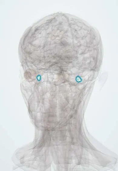
Ménière disease !
Ménière disease is an inner ear disorder with attacks of spinning vertigo, ringing, ear fullness and hearing loss that comes and goes.
Updated 2026-10-29
Structures involved
Tap one to see what it is and open it in 3D.
- Cochlea
- Vestibule
- Semicircular canals
- Vestibulocochlear nerve (VIII)
- Internal acoustic meatus
- Temporal bones
In histology
In the clinical exam
In physiology
- Vomiting and its control
- The vestibular system, tone and posture
- Hearing: middle ear, cochlea and auditory pathway
What it is
Inner ear disorder with recurrent attacks of vertigo, fluctuating sensorineural hearing loss, tinnitus and ear fullness, linked to endolymphatic hydrops (excess endolymph in the membranous labyrinth).
Who gets it
Usually begins between about 40 and 60 years of age. It is usually one-sided at first; the other ear can become involved over time.
Causes
- Idiopathic in most cases (Ménière disease)
- Secondary hydrops after trauma, infection or other inner ear disease (Ménière syndrome)
- Genetic and autoimmune factors proposed
Risk factors
- Family history
- Autoimmune disease
- Migraine (frequent association)
- Allergy (association proposed)
Pathophysiology
Endolymph is produced or absorbed abnormally, and the membranous labyrinth swells (endolymphatic hydrops). Changes in pressure and possible membrane ruptures disturb hair cell function in the cochlea and vestibule, causing attacks. Repeated attacks damage hair cells, leading to permanent, at first low-frequency, hearing loss.
Classification
- Definite Ménière disease
- Probable Ménière disease (diagnostic criteria vary between societies)
Symptoms and signs
- Spontaneous attacks of spinning vertigo lasting from about 20 minutes to 12 hours
- Fluctuating hearing loss in the affected ear, low frequencies first
- Tinnitus, often low-pitched and roaring
- Ear fullness or pressure
- Nausea and vomiting during attacks
- Sudden falls without loss of consciousness (Tumarkin drop attacks) in advanced cases
Diagnosis
- Clinical
- Clinical diagnosis based on accepted criteria: two or more spontaneous vertigo attacks of typical duration, audiometrically documented low- to mid-frequency sensorineural loss in the affected ear, and fluctuating aural symptoms, not better explained by another disorder.
- Tests
- Pure-tone audiometry is essential. Vestibular tests (caloric, video head impulse test), electrocochleography in selected cases.
- Imaging
- MRI with gadolinium of the internal acoustic meatus and brain to exclude vestibular schwannoma and other lesions; special delayed MRI sequences can show hydrops in some centres.
Differential diagnosis
- Vestibular migraine
- Vestibular schwannoma
- BPPV
- Vestibular neuritis or labyrinthitis
- Autoimmune inner ear disease
- Otosyphilis
Goal of treatment
Reduce the frequency and severity of vertigo attacks, preserve hearing where possible and maintain quality of life.
Treatment
- Lifestyle
- Lifestyle measures often advised: low-salt diet, limiting caffeine and alcohol, regular sleep and stress management; the evidence is limited.
- Acute
- During attacks, vestibular suppressants (antihistamines, benzodiazepines) and antiemetics for a short time.
- Medical
- Long-term medicines such as diuretics or betahistine are used in many countries; evidence of benefit is mixed and practice varies.
- Procedure
- Intratympanic injections: corticosteroids, or gentamicin to ablate vestibular function in disabling vertigo (risk to hearing).
- Surgical
- Surgery for refractory vertigo: endolymphatic sac surgery, vestibular nerve section, or labyrinthectomy when hearing is no longer useful.
- Rehabilitation
- Vestibular rehabilitation between attacks, and hearing aids or cochlear implant for hearing loss.
Complications
- Permanent sensorineural hearing loss
- Chronic tinnitus
- Involvement of the other ear
- Falls and drop attacks
- Anxiety and depression
Prognosis
The course is unpredictable, with clusters of attacks and remissions. Vertigo often lessens over the years, but hearing loss tends to progress and become permanent.
Test yourself
Think of the answer, then tap the question to see it.
What is the classic tetrad of Ménière disease?
Episodic vertigo, fluctuating sensorineural hearing loss, tinnitus and ear fullness.
What histopathological change underlies Ménière disease?
Endolymphatic hydrops: distension of the membranous labyrinth by excess endolymph.
Which frequencies are typically affected first on audiometry?
Low frequencies, in a fluctuating sensorineural pattern.
Why is MRI done in suspected Ménière disease?
To exclude a vestibular schwannoma or other lesion of the internal acoustic meatus or brain.
Which intratympanic drug can ablate vestibular function in refractory Ménière disease?
Gentamicin, which is vestibulotoxic; it carries a risk of hearing loss.
Related conditions
- Benign paroxysmal positional vertigo (BPPV)
- Vestibular schwannoma (acoustic neuroma)
- Migraine
- Presbycusis (age-related hearing loss)
Educational information, not medical advice. For any symptom, see a health professional.
Sources: Meniere Disease (StatPearls (NCBI Bookshelf), 2026).
Spotted a mistake?We are always improving for the community. Tell us what is wrong and our team will review it.Report a mistake
Representative model, for educational purposes only.