Adrenocortical carcinoma !
A rare, aggressive cancer of the adrenal cortex, often presenting with fast-onset Cushing syndrome or virilisation, or as a large adrenal mass.
Updated 2026-11-01
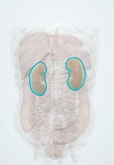
Structures involved
Tap one to see what it is and open it in 3D.
Why it happens
- Tumour suppressor genes: RB, TP53, APC, BRCA and the two-hit hypothesis
- Hereditary cancer syndromes: the table to know
- Adrenal cortisol synthesis
- Steroid hormone synthesis (overview)
- Adrenal cortex zones and medulla
What it is
A malignant tumour of the adrenal cortex. Most secrete hormones, especially cortisol and androgens, producing rapidly progressive Cushing syndrome, virilisation in women or feminisation in men. Others present as a large abdominal mass or are found on imaging.
Who gets it
Rare, about 1 to 2 cases per million people per year, with a peak in early childhood and another in the fourth to fifth decades; slightly more common in women. Southern Brazil has a childhood incidence 10 to 15 times the world rate because of the inherited TP53 R337H variant, a Latin American fact relevant across the region.
Causes
- Most adult cases are sporadic, with somatic TP53, CTNNB1 (beta-catenin) and other mutations
- Li-Fraumeni syndrome (germline TP53), the main cause in children
- TP53 R337H founder variant in southern Brazil
- Beckwith-Wiedemann syndrome, Lynch syndrome and MEN 1 in a minority
Risk factors
- Family history of early cancers (Li-Fraumeni pattern: sarcoma, breast, brain tumours, leukaemia)
- Childhood with a germline TP53 variant
- Beckwith-Wiedemann syndrome or Lynch syndrome
Pathophysiology
Malignant cortical cells often lose control of steroid synthesis, so they release cortisol together with androgen precursors (such as DHEA-S) and sometimes oestrogen, aldosterone or steroid intermediates. Mixed cortisol and androgen excess strongly suggests carcinoma. The tumour grows large, invades the kidney and inferior vena cava, and spreads to liver, lung and bone. Cortisol from the tumour suppresses ACTH, so the opposite adrenal atrophies, and adrenal insufficiency can follow surgery.
Classification
- Functioning (cortisol, androgens, oestrogen, aldosterone or mixed) versus non-functioning
- ENSAT stages I–II (localised, up to 5 cm or larger), III (local invasion, nodes or venous tumour thrombus), IV (distant metastases)
- Weiss score on histology distinguishes carcinoma from adenoma; Ki-67 index grades it
Symptoms and signs
- Rapid weight gain, moon face, muscle weakness, bruising, hypertension and hyperglycaemia (Cushing syndrome)
- Hirsutism, deep voice, clitoromegaly, acne and amenorrhoea in women; precocious puberty in children
- Gynaecomastia and testicular atrophy in men (oestrogen-secreting tumours)
- Flank or abdominal pain, fullness or a palpable mass
- Hypokalaemia and hypertension; fever, weight loss and leg swelling in advanced disease
Diagnosis
- Clinical
- Speed of onset and combination of hormone features, family history of early cancers, examination for virilisation and abdominal mass.
- Tests
- Full hormone work-up before surgery: dexamethasone suppression test, ACTH, DHEA-S, testosterone, androstenedione, 17-hydroxyprogesterone, oestradiol in men, aldosterone and renin with potassium in hypertensive patients, and metanephrines to exclude pheochromocytoma. Germline TP53 testing in all children and in adults with suggestive family history. Biopsy is avoided when carcinoma is suspected because of tumour seeding, unless needed for metastatic disease.
- Imaging
- CT or MRI shows a large, heterogeneous mass with necrosis, calcification, high unenhanced attenuation (over 10 Hounsfield units) and slow contrast washout; chest CT and FDG-PET for staging and venous invasion.
Differential diagnosis
- Benign adrenocortical adenoma
- Pheochromocytoma
- Adrenal metastasis (lung, kidney, melanoma)
- Renal cell carcinoma invading the adrenal
- Androgen excess from polycystic ovary syndrome, ovarian tumours or non-classic congenital adrenal hyperplasia
Goal of treatment
Complete removal of the tumour with an intact capsule by an experienced surgeon, control of hormone excess, protection against post-operative adrenal insufficiency, and adjuvant or systemic therapy based on recurrence risk.
Treatment
- Surgical
- Open radical adrenalectomy with en bloc removal of invaded structures for stage I–III disease; tumour rupture must be avoided. Resection of selected metastases or recurrences.
- Medical
- Mitotane (an adrenolytic agent) as adjuvant therapy in high-risk patients and for advanced disease, often combined with platinum-based chemotherapy; glucocorticoid replacement is needed during mitotane therapy.
- Medical
- Steroidogenesis inhibitors to control severe cortisol excess.
- Procedure
- Radiotherapy to the tumour bed in selected cases and for painful bone metastases.
- Follow-up
- Imaging of chest and abdomen and hormone markers every few months for years; mitotane blood levels; cortisol and potassium after surgery.
Complications
- Local recurrence and metastases to liver, lung and bone
- Inferior vena cava invasion and thromboembolism
- Adrenal insufficiency after surgery or during mitotane therapy
- Complications of cortisol excess: infections, diabetes, hypertension, osteoporosis
- Hypokalaemia
Prognosis
Poor overall: about 40% of patients are alive at five years, falling to under 20% with distant metastases. Complete resection at an early stage and a low Ki-67 index carry the best outlook; children, particularly with small tumours, do better than adults.
Test yourself
Think of the answer, then tap the question to see it.
Which hormonal pattern most strongly suggests adrenocortical carcinoma rather than adenoma?
Combined cortisol and androgen (for example, DHEA-S) excess, or virilisation developing quickly.
Why is percutaneous biopsy avoided when adrenocortical carcinoma is suspected?
It can seed tumour along the needle track and rarely changes surgical management; pheochromocytoma must also be excluded first.
Which hereditary syndrome is the main cause of adrenocortical carcinoma in children?
Li-Fraumeni syndrome (germline TP53), including the R337H variant common in southern Brazil.
What is the adrenolytic drug used after surgery in high-risk adrenocortical carcinoma?
Mitotane, with glucocorticoid replacement during treatment.
Which CT features of an adrenal mass favour malignancy?
Size over 4 cm, heterogeneity, unenhanced attenuation above 10 HU and slow contrast washout.
Related conditions
- Adrenal incidentaloma
- Cushing syndrome
- Primary hyperaldosteronism (Conn syndrome)
- Phaeochromocytoma
- Adrenal insufficiency
- Multiple endocrine neoplasia (MEN 1, 2A, 2B)
- Hypokalaemia
- Polycystic ovary syndrome
- Renal cell carcinoma
- Type 2 diabetes mellitus
- Arterial hypertension
Educational information, not medical advice. For any symptom, see a health professional.
Sources: Adrenocortical Carcinoma Treatment (PDQ®)–Health Professional Version (National Cancer Institute (PDQ), 2025); An inherited p53 mutation that contributes in a tissue-specific manner to pediatric adrenal cortical carcinoma (Proceedings of the National Academy of Sciences of the USA (PubMed), 2001).
Spotted a mistake?We are always improving for the community. Tell us what is wrong and our team will review it.Report a mistake
Representative model, for educational purposes only.