Conditions · Infectious and tropical diseases
Aspergillosis !
Aspergillosis is a group of lung and sinus diseases caused by a common mould: allergic, a fungus ball in old cavities, or invasive in weak immunity.
Updated 2026-11-01
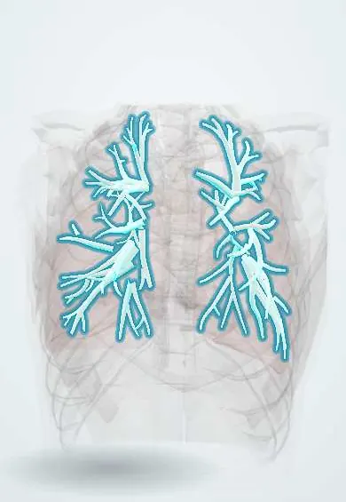
Structures involved
Tap one to see what it is and open it in 3D.
- Segmental bronchi
- Nasal cavity
- Lobar bronchi
- Main bronchi
- Pleura
- Maxillary sinus
- Ethmoid cells
- Sphenoid sinus
Microorganisms that cause it
Why it happens
- Infarction: red vs white, and what decides the damage
- White blood cells: types and functions
- The airway: from nose to bronchioles
What it is
Disease caused by inhaling spores of Aspergillus, a mould found everywhere in soil, compost and dust. Depending on the host it causes allergic disease (allergic bronchopulmonary aspergillosis), colonisation of old cavities (aspergilloma), chronic pulmonary aspergillosis or life-threatening invasive aspergillosis. Mucormycosis, a related angioinvasive mould infection, is covered in a section below.
Who gets it
Exposure is universal, but disease depends on the lungs and immunity. In Colombia and Latin America, chronic pulmonary aspergillosis is linked to cavities left by tuberculosis, while invasive disease appears in patients with leukaemia, neutropenia, transplants, high-dose corticosteroids and, more recently, severe influenza or COVID-19 in intensive care.
Causes
- Aspergillus fumigatus (most common), A. flavus, A. niger, A. terreus
- Inhalation of airborne conidia; hospital outbreaks linked to construction or contaminated air
- Mucormycosis: Rhizopus, Mucor and related moulds (section below)
Risk factors
- Prolonged neutropenia (acute leukaemia, chemotherapy, stem cell transplant)
- Solid organ transplant and high-dose corticosteroids
- Asthma and cystic fibrosis (allergic form)
- Old lung cavities from tuberculosis, sarcoidosis or bronchiectasis (aspergilloma and chronic form)
- COPD, severe influenza or COVID-19 in intensive care
- Uncontrolled diabetes with ketoacidosis and iron overload (mucormycosis)
Pathophysiology
Normally alveolar macrophages and neutrophils clear inhaled conidia. In atopic airways the fungus provokes an IgE- and eosinophil-driven reaction with mucus plugging and central bronchiectasis (ABPA). In pre-existing cavities it grows as a ball of hyphae. When neutrophils are absent, hyphae invade blood vessels, causing infarction, haemorrhage and spread to brain and other organs. Aspergillus hyphae are septate and branch at acute angles; Mucorales hyphae are broad, ribbon-like and branch at right angles.
Classification
- Allergic bronchopulmonary aspergillosis (ABPA) and allergic fungal sinusitis
- Aspergilloma (fungus ball) in a pre-existing cavity
- Chronic pulmonary aspergillosis (chronic cavitary or fibrosing)
- Invasive pulmonary aspergillosis and disseminated disease
- Invasive sinus disease
- Mucormycosis: rhino-orbito-cerebral, pulmonary, cutaneous
Symptoms and signs
- ABPA: poorly controlled asthma, wheeze, brown mucus plugs, recurrent infiltrates
- Aspergilloma: often silent, or recurrent haemoptysis
- Chronic form: months of cough, weight loss, fatigue and haemoptysis
- Invasive form: fever despite antibiotics in a neutropenic patient, cough, pleuritic chest pain and haemoptysis
- Sinus disease: facial pain, nasal congestion and discharge
- Mucormycosis: facial swelling, black nasal or palatal eschar, eye swelling and loss of vision
Diagnosis
- Clinical
- Depends on the form: an asthmatic with worsening control and eosinophilia (ABPA), a cavity with haemoptysis (aspergilloma), or a febrile neutropenic patient with new lung nodules (invasive).
- Tests
- ABPA: very high total IgE, Aspergillus-specific IgE and IgG, eosinophilia and positive skin test. Invasive: galactomannan in serum or bronchoalveolar lavage, beta-D-glucan, culture and PCR; histology showing tissue invasion confirms it. Chronic form and aspergilloma: Aspergillus IgG. Mucormycosis: tissue biopsy for broad non-septate hyphae.
- Imaging
- Chest CT: nodules with a halo sign early and an air-crescent sign later in invasive disease; a mobile ball within a cavity in aspergilloma; central bronchiectasis and mucus plugs in ABPA. Sinus and orbit CT or MRI for sinus disease and mucormycosis.
Differential diagnosis
- Pulmonary tuberculosis and its reactivation
- Bacterial pneumonia and lung abscess
- Lung cancer
- Eosinophilic pneumonia and eosinophilic granulomatosis with polyangiitis
- Pneumocystis pneumonia in immunosuppressed patients
Goal of treatment
Match therapy to the form: calm the allergic reaction in ABPA, stop bleeding and remove fungus balls when needed, and start antifungals early in invasive disease while restoring immunity.
Treatment
- Medical
- ABPA: systemic corticosteroids to control the allergic reaction, often with an oral azole antifungal to reduce fungal load and steroid needs.
- Medical
- Invasive and chronic aspergillosis: triazole antifungals (voriconazole or isavuconazole first line), with lipid amphotericin B or echinocandins as alternatives; reduction of immunosuppression where possible. Mucormycosis needs amphotericin B, not voriconazole.
- Surgical
- Resection of a symptomatic aspergilloma or localised disease; urgent, extensive debridement of necrotic tissue in mucormycosis and invasive sinus disease.
- Procedure
- Bronchial artery embolisation for massive or recurrent haemoptysis.
- Follow-up
- Galactomannan screening and CT in high-risk haematology patients, antifungal prophylaxis in selected cases, protective environment with filtered air, and glucose control in diabetes.
Drugs for this condition
Complications
- Massive haemoptysis
- Respiratory failure
- Spread to brain, skin, heart or other organs
- Central bronchiectasis and pulmonary fibrosis (ABPA)
- Orbital and cavernous sinus involvement (mucormycosis)
Prognosis
ABPA is controllable but tends to relapse. Aspergilloma may stay stable for years. Invasive aspergillosis has high mortality, about 30–60%, and improves with early antifungals and recovery of neutrophils; mucormycosis is often fatal without combined surgery and amphotericin B.
Prevention
- Filtered air and control of construction dust in units with immunosuppressed patients
- Antifungal prophylaxis for selected high-risk haematology patients
- Good control of asthma, cystic fibrosis and diabetes
Test yourself
Think of the answer, then tap the question to see it.
Which patients develop allergic bronchopulmonary aspergillosis?
People with asthma or cystic fibrosis.
What is the typical CT sign of early invasive pulmonary aspergillosis?
A nodule with a surrounding halo of ground-glass opacity (halo sign).
How do Aspergillus hyphae look under the microscope?
Septate hyphae branching at acute angles (about 45 degrees).
What is the first-line antifungal class for invasive aspergillosis?
Triazoles, such as voriconazole or isavuconazole.
Which patient typically gets rhino-orbito-cerebral mucormycosis?
A person with uncontrolled diabetes and ketoacidosis, or severe neutropenia.
Related conditions
- Asthma
- Cystic fibrosis
- Bronchiectasis
- Pulmonary tuberculosis
- Febrile neutropenia
- Acute leukaemia (AML and ALL)
- Chronic obstructive pulmonary disease (COPD)
- Chronic rhinosinusitis and nasal polyps
- Candidiasis: oral, oesophageal and invasive
- Endemic mycoses: blastomycosis and coccidioidomycosis
- Lung cancer
- Pneumocystis pneumonia (PCP)
Educational information, not medical advice. For any symptom, see a health professional.
Sources: Aspergillosis Basics | Aspergillosis | CDC (Centers for Disease Control and Prevention, 2024); Aspergillosis: MedlinePlus Medical Encyclopedia (MedlinePlus (U.S. National Library of Medicine), 2024); Mucormycosis Basics | Mucormycosis | CDC (Centers for Disease Control and Prevention, 2024).
Spotted a mistake?We are always improving for the community. Tell us what is wrong and our team will review it.Report a mistake
Representative model, for educational purposes only.